
Page 198 - Veterinary Histology of Domestic Mammals and Birds, 5th Edition
P. 198
180 Veterinary Histology of Domestic Mammals and Birds
sensory receptors. Intra-epithelial Langerhans cells func- Lip (labium)
VetBooks.ir tion as immune receptor cells in local immune responses. The upper and lower lips surround the entrance to the oral
Differentiation of the mucosa of the oral cavity is evi-
cavity. They are used for sucking and prehension and may
dent in the gums (gingiva). In this region, the mucosa have a tactile function. The lips (Figure 10.2) are composed
covers the alveolar processes of the maxilla and mandi- of the following layers:
ble. The stratified squamous epithelium is keratinised to
some extent and exhibits rapid regeneration. Taut bands · outer layer of skin:
of collagen fibres bind the tela submucosa tightly to the − modified epidermis,
periosteum of the underlying bone. − sensory receptors and tactile corpuscles,
The lamina propria is composed of loose connective − individually distinct surface patterns (planum
tissue containing a network of collagen and elastic fibres. nasolabiale),
Abundant vessels and nerves are present. Papillae that − subepithelial serous glands (planum nasolabiale,
interdigitate with the epithelium are particularly promi- planum rostrale),
nent in the palate and tongue, reflecting the mechanical · muscle layer
load borne by these structures. This feature serves to − m. orbicularis oris
anchor, and facilitate nourishment of, the overlying epi- · internal mucosal layer:
thelium. The mucosa (epithelium and lamina propria) is − non-glandular mucosa and
non-glandular (tunica mucosa non-glandularis). − papillae, glands
Lying deep to the non-glandular mucosa is a layer of
connective tissue termed the tela submucosa. This layer The external surface of the lips consists of modified skin
contains blood vessels, a well-developed submucosal nerve (integumentum commune). Sinus hairs are present in
plexus (Meissner’s plexus, plexus nervorum submucosus), carnivores, small ruminants and horses (see Chapter 15,
regionally distributed serous, mucous or mixed glands ‘Common integument’). Species-specific modifications
(e.g. labial, buccal and lingual glands) and aggregates of the skin of the upper lip include the planum rostrale of
of lymphatic tissue (lymphoid follicles and tonsils). In pigs, the planum nasolabiale of cattle and the philtrum of
locations subjected to increased shear forces, the tela sub- carnivores and small ruminants. The planum nasolabiale
mucosa is particularly taut, relatively immobile and tightly and planum rostrale lack sebaceous glands and hair (occa-
bound to the underlying periosteum. Glands are absent in sional hairs are found on the planum rostrale). Superficially,
the tip and dorsum of the tongue, the hard palate and the the lips are richly endowed with free sensory nerve end-
gums. The wall of the oral cavity is extensively reinforced ings. Particularly in pigs, the dermis contains numerous
by the presence of skeletal muscle in the tunica muscula- encapsulated tactile corpuscles. The stratified squamous
ris (lips, cheeks and soft palate). Skeletal muscle also forms epithelium of the outer surface of the lips interdigitates
the structural foundation of the tongue (for further infor- with pronounced connective tissue papillae. In cattle this
mation regarding the layers of the wall of the digestive results in grooves that form individually distinctive pat-
tract see ‘Structure of tubular digestive organs’). terns on the planum nasolabiale (Figure 10.3).
Liver Pancreas
Oral cavity
Colon
Pharynx Caecum
Rectum
Anal canal
Oesophagus
Stomach
Duodenum Jejunum Ileum
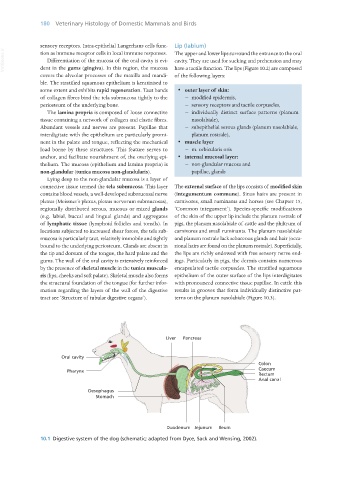
10.1 Digestive system of the dog (schematic; adapted from Dyce, Sack and Wensing, 2002).
Vet Histology.indb 180 16/07/2019 15:00