
Page 205 - Veterinary Histology of Domestic Mammals and Birds, 5th Edition
P. 205
Digestive system (apparatus digestorius) 187
by the mandibular branch of the trigeminal nerve (V) and (pulpa dentis). Towards the proximal end of the tooth, the
VetBooks.ir the facial nerve (VII), glossopharyngeal nerve (IX) and pulp cavity narrows to become the root canal (canalis radi-
cis dentis) that ends in the apical foramen (foramen apicale
vagus (X) nerve.
Blood supply to the tongue comprises dense capillary dentis) (Figure 10.12). The apical foramen is traversed by
networks that tend to accumulate in subepithelial sheets. nerves and blood vessels entering and leaving the pulp.
The margins of the tongue are particularly well vascular- In this region, odontoblasts (see below) produce pre-
ised. Mixed glands lie along the lateral margins of the dentin and contribute to the mineralisation of the tooth
tongue in horses and cattle. root (root dentin). External to the dentin is a bone-like
substance, termed cementum, that is produced through-
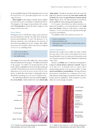
Tooth (dens) out life by cementoblasts.
Working in concert with the lips, tongue, upper and lower The crown consists, from exterior to interior, of:
jaw and masticatory muscles, the teeth serve in the pre-
hension and breakdown of food. The dentition of the · enamel (enamelum),
domestic mammals varies with species, the structure of · dentin (dentinum) and
the teeth being influenced by diet. Despite these differ- · pulp (pulpa coronalis) within the pulp cavity (cavum
ences, teeth are consistent in their basic structure (Figures coronalis dentis).
10.10 to 10.12), consisting of the:
Enamel (enamelum)
· crown (corona dentis), Enamel is the hardest tissue within the body. It forms
· neck (cervix dentis) and the outer layer of the crown of brachydont teeth and is
· root (radix dentis). located beneath a layer of cementum in hypsodont teeth
(see Veterinary Anatomy of Domestic Mammals: Textbook and
The crown of the tooth is the visible (free, distal) portion Colour Atlas).
that protrudes beyond the gingiva. The neck is surrounded Enamel is acellular and is composed of hexagonal to
by the gingiva. The root is the proximal portion of the polygonal enamel prisms (prismata enameli) (Figures
tooth, which is embedded in the alveoli of the maxilla and 10.10 and 10.11). It develops from an enamel matrix
mandible. These divisions are obvious in brachydont teeth. formed embryonically as the secretory product of ecto-
Hypsodont teeth (teeth of horses, cheek teeth of rumi- dermal ameloblasts (enameloblasts). During embryonic
nants), in which the neck is hard to distinguish, may be development, ameloblasts are a component of the exter-
described as consisting of a root and an elongated body. nally oriented enamel organ and spread to form a sheet of
The mineralised wall of the tooth surrounds the pulp cav- simple columnar epithelium. Their cytoplasm is eosino-
ity (cavum dentis), which encloses the central dental pulp philic, the nuclei strongly basophilic.
10.10 Tooth of a calf during embryonic development. 10.11 Wall of the tooth of a calf during embryonic
Haematoxylin and eosin stain (x14). development. Haematoxylin and eosin stain (x120).
Vet Histology.indb 187 16/07/2019 15:00