
Page 248 - Veterinary Histology of Domestic Mammals and Birds, 5th Edition
P. 248
230 Veterinary Histology of Domestic Mammals and Birds
VetBooks.ir
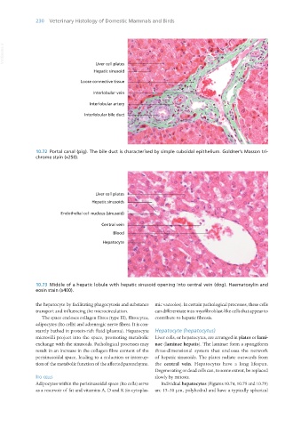
10.72 Portal canal (pig). The bile duct is characterised by simple cuboidal epithelium. Goldner’s Masson tri-
chrome stain (x250).
10.73 Middle of a hepatic lobule with hepatic sinusoid opening into central vein (dog). Haematoxylin and
eosin stain (x400).
the hepatocyte by facilitating phagocytosis and substance mic vacuoles). In certain pathological processes, these cells
transport and influencing the microcirculation. can differentiate into myofibroblast-like cells that appear to
The space encloses collagen fibres (type III), fibrocytes, contribute to hepatic fibrosis.
adipocytes (Ito cells) and adrenergic nerve fibres. It is con-
stantly bathed in protein-rich fluid (plasma). Hepatocyte Hepatocyte (hepatocytus)
microvilli project into the space, promoting metabolic Liver cells, or hepatocytes, are arranged in plates or lami-
exchange with the sinusoids. Pathological processes may nae (laminae hepatis). The laminae form a spongiform
result in an increase in the collagen fibre content of the three-dimensional system that encloses the network
perisinusoidal space, leading to a reduction or interrup- of hepatic sinusoids. The plates radiate outwards from
tion of the metabolic function of the affected parenchyma. the central vein. Hepatocytes have a long lifespan.
Degenerating or dead cells can, to some extent, be replaced
ito cells slowly by mitosis.
Adipocytes within the perisinusoidal space (Ito cells) serve Individual hepatocytes (Figures 10.74, 10.75 and 10.79)
as a reservoir of fat and vitamins A, D and K (in cytoplas- are 15–30 μm, polyhedral and have a typically spherical
Vet Histology.indb 230 16/07/2019 15:02