
Page 261 - Veterinary Histology of Domestic Mammals and Birds, 5th Edition
P. 261
Respiratory system (apparatus respiratorius) 243
The olfactory mucosa covers the ethmoturbinates, dorsal secretory product of these cells has a cleansing function,
VetBooks.ir endoturbinates and part of the caudal segment of the nasal facilitating the detection of subsequent olfactory stimuli.
septum. Modified bipolar neurons serve as neurosensory
Species variation
cells that detect odours (Figure 11.4). Axons leave the basal
portion of the cell forming bundles that pass through the Birds: The cranial portion of the nasal cavity (nasal
submucosa (see description of olfactory epithelium in vestibule) is lined with non-glandular mucosa.
Chapter 16, ‘Receptors and sense organs’). Caudally, in the respiratory region, this transitions to a
The lamina propria houses branched tubulo-acinar ciliated pseudostratified epithelium with secretory cells
olfactory glands (glandulae olfactoriae). The thin, watery (Figures 11.8 and 11.9). This is continued in the olfac-
glandular secretion contains enzymes (proteases) that tory region by olfactory epithelium. The histological
are important for breaking down odoriferous substances structure of the epithelium differs little from that of
and binding them to neurosensory cells. In addition, the mammals. Typically yellowish in colour, the olfactory
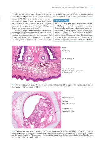
11.5 Vomeronasal organ (cat). The paired vomeronasal organ lies at the base of the osseous nasal septum.
Haematoxylin and eosin stain (x15).
11.6 Vomeronasal organ (calf). The interior of the vomeronasal organ is lined medially by olfactory mucosa and
laterally by respiratory mucosa. Glandular aggregates and expanded veins lie between the respiratory mucosa
and the external cartilage. The respiratory mucosa contains poorly myelinated subepithelial nerve fibre bundles.
Haematoxylin and eosin stain (x40).
Vet Histology.indb 243 16/07/2019 15:02