
Page 266 - Veterinary Histology of Domestic Mammals and Birds, 5th Edition
P. 266
248 Veterinary Histology of Domestic Mammals and Birds
VetBooks.ir
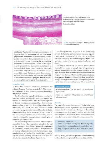
11.14 Trachea (chicken). Haematoxylin
and eosin stain (x70).
semilunaris. Together, the cartilaginous components of The intra-pulmonary segments of the conducting
the syrinx form the tympanum. Left and right lateral airways, the bronchi and bronchioles, represent approxi-
tympaniform membranes (membrana tympaniformis mately 6% of the volume of the lung. Over 85% of lung
lateralis) extend from the tympanum to the lateral side volume is formed by the respiratory parenchyma – the
of the bronchial cartilages. Paired medial tympaniform respiratory bronchioles, alveolar ducts, alveolar sacs and
membranes (membrana tympaniformis medialis) pass alveoli.
from the pessulus to the (incomplete) medial aspect of The lung is enclosed by the visceral pleura (pleura
the bronchial cartilages. Elastic connective tissue pads visceralis), composed of a single layer of squamous to
termed labia project from the membranes into the cuboidal mesothelial cells and a variably thick layer of col-
lumen of the syrinx. During phonation, the membranes lagen and elastic fibres. Connective tissue septa extend from
and labia function in a similar manner to the vocal folds the pleura into the lung. This interstitial connective tissue
of the mammalian larynx. Syringeal muscles are pre- (interstitium) divides the lobes of the lung into lobules.
sent in song birds and absent in domestic poultry. Intra-pulmonary vessels and nerves accompanying the con-
nective tissue septa account for around 10% of lung volume.
Lung (pulmo)
At the tracheal bifurcation, the trachea divides into two Species variation
primary bronchi (bronchi principales). The primary Ruminants and pig: The pulmonary interstitial tissue
bronchi branch to form the intra-pulmonary lobar bron- is prominent.
chi (bronchi lobares). Horse and carnivores: The pulmonary interstitium is
The lobar bronchi typically divide into two further relatively sparse.
branches, each of which divides again (into bronchi seg-
mentale and subsegmentales). This dichotomous process
of division continues, accompanied by a decrease in the Bronchi
diameter of the airway, until the small bronchioles (bron- The main differences in the structure of the bronchi (bron-
chioli veri) are formed. The small bronchioles divide chi principales, lobares, segmentales and subsegmentales),
extensively, giving rise to the terminal bronchioles (bron- compared with the trachea, are the shape of the cartilagi-
chioli terminales). These represent the final portion of nous elements and the arrangement of the smooth muscle
the conducting component of the respiratory system, fibres (Figures 11.16 to 11.19). From innermost to outer-
which is continued by the respiratory portion (region of most, the bronchi consist of the following components:
gas exchange) (Figure 11.15).
The respiratory portion commences with the appear- · respiratory epithelium,
ance of the first sac-like evaginations (alveoli) in the wall · lamina propria with mixed glands (bronchial glands),
of the respiratory bronchioles (bronchioli respiratorii · elastic fibre network and smooth muscle layer and
or alveolares). Further divisions result in the formation · rings of hyaline cartilage with associated elastic
of narrow tubular airways termed alveolar ducts (duc- fibres.
tus alveolares). The walls of the alveolar ducts consist of
adjacent alveolar openings. The alveolar ducts open into Distally, the height of the respiratory epithelium lining
alveolar sacs (sacculi alveolares) (Figure 11.16). the bronchial lumen gradually decreases (Figure 11.18).
Vet Histology.indb 248 16/07/2019 15:03