Page 269 - Veterinary Histology of Domestic Mammals and Birds, 5th Edition
P. 269
Respiratory system (apparatus respiratorius)   251


       VetBooks.ir


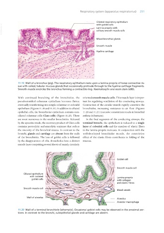
                  11.19  Wall of a bronchus (pig). The respiratory epithelium rests upon a lamina propria of loose connective tis-
                  sue with coiled, tubular mucous glands that occasionally protrude through to the hyaline cartilage fragments.
                  Smooth muscle encircles the bronchus forming a contractile ring. Haematoxylin and eosin stain (x80).


                  With continued branching of the bronchioles, the  oriented smooth muscle cells. This muscle layer is impor-
                  pseudostratified columnar epithelium becomes flatter,  tant for regulating ventilation of the conducting airways.
                  eventually transforming into simple columnar or cuboidal  Contraction of the circular muscle rapidly constricts the
                  epithelium (Figures 11.20 and 11.21). In addition to ciliated  bronchioles, increasing resistance to air flow (Figures
                  epithelial cells, the bronchiolar epithelium contains non-  11.20 and 11.21) (excessive constriction results in bronchial
                  ciliated columnar cells (Clara cells) (Figure 11.25). These  asthma in humans).
                  are most numerous in the smaller bronchioles. Released   In the final segments of the conducting airways, the
                  by the apocrine mode, the secretory product of Clara cells  terminal bronchi, the epithelium is reduced to a single
                  contains proteolytic and mucolytic enzymes that reduce  layer of cuboidal cells and the number of elastic fibres
                  the viscosity of the bronchial mucus. In contrast to the  in the lamina propria increases. In conjunction with the
                  bronchi, glands and cartilage are absent from the walls  well-developed bronchiolar muscle, the constrictive
                  of the bronchioles. The loss of goblet cells is followed  effect of the elastic fibres contributes to folding of the
                  by the disappearance of cilia. Bronchioles have a distinct  mucosa.
                  muscle layer comprising several sheets of mainly circularly


                  11.20  Wall of a terminal bronchiole (schematic). Occasional goblet cells may be observed in the proximal por-
                  tions. In contrast to the bronchi, subepithelial glands and cartilage are absent.


       Vet Histology.indb   251                                                                                  16/07/2019   15:03

