Page 274 - Veterinary Histology of Domestic Mammals and Birds, 5th Edition
P. 274
256  Veterinary Histology of Domestic Mammals and Birds


                                                                  tubules. In most species their diameter is around 0.5 mm
                     ·  numerous laterodorsal secondary bronchi,
       VetBooks.ir   ·  7–10 mediodorsal secondary bronchi,       (1–1.5 mm in the chicken).
                                                                     Parabronchi have several distinctive features (Figures
                     ·  4–7 lateroventral secondary bronchi and
                                                                  11.28 and 11.29):
                     ·  4 medioventral secondary bronchi.

                   Third-order bronchi, or parabronchi, are also referred to   ·  they anastomose with one another,
                   as ‘air pipes’. Parabronchi arising from mediodorsal sec-  ·  chambers called atria open from their walls into
                   ondary bronchi within the dorsal palaeopulmo meet and   infundibula and air capillaries,
                   anastomose with their ventral counterparts in the inte-  ·  they contain gas exchange units,
                   rior of the lung (refer to anatomy texts). The parabronchi   ·  their diameter is uniform within species and
                   are arranged in a parallel hexagonal array of elongated   ·  they manifest as hexagonal tubes.

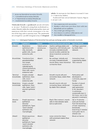
                   Table 11.2  Histological features of the bronchial tree and gas exchange system of domestic mammals.

                    Segment     Epithelium        Glands         Connective tissue and muscle  Special features
                    Bronchi     Respiratory       Tubulo-acinar   Hyaline cartilage plates and   Cartilage supported
                                epithelium with   seromucous     fragments, with helically and   by elastic fibre
                                cilia, microvilli,   glands      circularly oriented smooth   network
                                goblet cells, neuro-             muscle bundles underlain by
                                endocrine cells                  dense interstitial connective
                                                                 tissue
                    Bronchioles  Transforms from   Absent        No cartilage, helically and   Connective tissue
                                pseudostratified                 circularly oriented smooth   supported by elastic
                                into simple                      muscle fibres, loose interstitial  fibre network
                                columnar or                      connective tissue
                                cuboidal partly
                                ciliated epithelium,
                                Clara cells, neuro-
                                endocrine cells
                    Terminal    Simple cuboidal   Absent         Smooth muscle cells and    Particularly well
                    bronchioles  epithelium, Clara               connective tissue, elastic fibre   developed in
                                cells, neuro-                    network                    ruminants, pigs and
                                endocrine cells                                             horses
                    Respiratory   Simple cuboidal   Absent       Smooth muscle cells and    Number of alveoli
                    bronchioles  epithelium, Clara               connective tissue, reinforced   increases with
                                cells, Types I and II            by elastic fibre network   each generation
                                alveolar epithelial                                         (I–III), abundant
                                cells                                                       in carnivores,
                                                                                            occasionally seen in
                                                                                            the horse, rare in the
                                                                                            pig and in ruminants
                    Alveolar    Simple squamous   Absent         Elastic fibre network, muscle   Up to 10 alveolar
                    ducts       epithelium                       cells in inter-alveolar septa   ducts are formed
                                (alveolar                        (acts as sphincter)        from the branching
                                epithelium), Types                                          of each respiratory
                                I and II alveolar                                           bronchiole,
                                epithelial cells,                                           branching of alveolar
                                macrophages                                                 ducts gives rise to
                                                                                            alveolar sacs
                    Alveoli     Simple squamous   Absent         Loose connective tissue    Blood–air barrier
                                epithelium                       with prominent elastic fibre
                                (alveolar                        network
                                epithelium), Types
                                I and II alveolar
                                epithelial cells,
                                macrophages


       Vet Histology.indb   256                                                                                  16/07/2019   15:03

