
Page 278 - Veterinary Histology of Domestic Mammals and Birds, 5th Edition
P. 278
260 Veterinary Histology of Domestic Mammals and Birds
VetBooks.ir
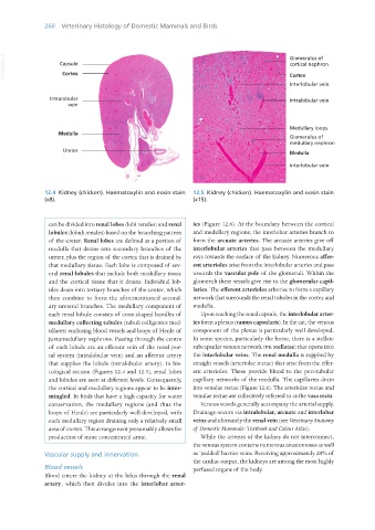
12.4 Kidney (chicken). Haematoxylin and eosin stain 12.5 Kidney (chicken). Haematoxylin and eosin stain
(x8). (x15).
can be divided into renal lobes (lobi renales) and renal ies (Figure 12.6). At the boundary between the cortical
lobules (lobuli renales) based on the branching pattern and medullary regions, the interlobar arteries branch to
of the ureter. Renal lobes are defined as a portion of form the arcuate arteries. The arcuate arteries give off
medulla that drains into secondary branches of the interlobular arteries that pass between the medullary
ureter, plus the region of the cortex that is drained by rays towards the surface of the kidney. Numerous affer-
that medullary tissue. Each lobe is composed of sev- ent arterioles arise from the interlobular arteries and pass
eral renal lobules that include both medullary tissue towards the vascular pole of the glomeruli. Within the
and the cortical tissue that it drains. Individual lob- glomeruli these vessels give rise to the glomerular capil-
ules drain into tertiary branches of the ureter, which laries. The efferent arterioles arborise to form a capillary
then combine to form the aforementioned second- network that surrounds the renal tubules in the cortex and
ary ureteral branches. The medullary component of medulla.
each renal lobule consists of cone-shaped bundles of Upon reaching the renal capsule, the interlobular arter-
medullary collecting tubules (tubuli colligentes med- ies form a plexus (ramus capsularis). In the cat, the venous
ullares) enclosing blood vessels and loops of Henle of component of the plexus is particularly well developed.
juxtamedullary nephrons. Passing through the centre In some species, particularly the horse, there is a stellate
of each lobule are an efferent vein of the renal por- subcapsular venous network (vv. stellatae) that opens into
tal system (intralobular vein) and an afferent artery the interlobular veins. The renal medulla is supplied by
that supplies the lobule (intralobular artery). In his- straight vessels (arteriolae rectae) that arise from the effer-
tological section (Figures 12.4 and 12.5), renal lobes ent arterioles. These provide blood to the peri-tubular
and lobules are seen at different levels. Consequently, capillary networks of the medulla. The capillaries drain
the cortical and medullary regions appear to be inter- into venulae rectae (Figure 12.6). The arteriolae rectae and
mingled. In birds that have a high capacity for water venulae rectae are collectively referred to as the vasa recta.
conservation, the medullary regions (and thus the Venous vessels generally accompany the arterial supply.
loops of Henle) are particularly well developed, with Drainage occurs via intralobular, arcuate and interlobar
each medullary region draining only a relatively small veins and ultimately the renal vein (see Veterinary Anatomy
area of cortex. This arrangement presumably allows for of Domestic Mammals: Textbook and Colour Atlas).
production of more concentrated urine. While the arteries of the kidney do not interconnect,
the venous system contains numerous anastomoses as well
Vascular supply and innervation as ‘padded’ barrier veins. Receiving approximately 20% of
the cardiac output, the kidneys are among the most highly
Blood vessels perfused organs of the body.
Blood enters the kidney at the hilus through the renal
artery, which then divides into the interlobar arter-
Vet Histology.indb 260 16/07/2019 15:03