
Page 280 - Veterinary Histology of Domestic Mammals and Birds, 5th Edition
P. 280
262 Veterinary Histology of Domestic Mammals and Birds
Species variation demonstrated through kidney transplantation that transec-
VetBooks.ir Birds: Branching of the intralobular vessels resembles Microscopic structure of the nephron and
tion of nerve fibres does not impact on kidney function.
that of mammals. The intralobular artery gives off
afferent arterioles (arteriolae glomerulares afferentes).
Post-glomerular efferent arterioles (arteriolae glomeru- collecting duct system
lares efferentes) give rise to capillaries that anastomose The functional unit of the kidney, consisting of the neph-
with the capillaries of the renal portal system, together ron and collecting duct system, is structurally similar in all
forming the peri-tubular capillary network that sur- domestic mammals. The nephron and collecting system are
rounds the tubules of the nephron (Figure 12.7). connected in series. Together with the renal interstitium and
the peri-tubular capillary network, they serve to produce and
modify the urine. The number of functional units per kidney
Lymph vessels varies with species (approximate number of nephrons per
The lymph vessels travel together with the arteries and kidney: horse – 2.7 million, ox – 4 million, small ruminants –
veins. In the capsule, they form a superficial drainage 0.5 million, pig – 1 million, dog – 180,000 to 400,000).
system. Embryologically, the nephron develops from the
metanephrogenic blastema. Under the influence of the
Nerves ureteric bud and its derivatives, the metanephrogenic
The kidney, particularly the blood vasculature, is inner- blastema transforms into a tube. The blind end of each
vated by numerous unmyelinated sympathetic nerve developing nephron undergoes balloon-like expansion,
fibres originating from the coeliac plexus. Some branches later differentiating into the bi-layered Bowman’s capsule.
extend through the interstitium to the juxtaglomerular This component of the nephron is invaginated by a capil-
apparatuses. Occasionally, smaller nerve fibre bundles are lary bundle (glomerulus). The inner (visceral) layer of the
closely apposed to the walls of renal tubules. Visceral affer- nephron-anlage becomes closely associated with these cap-
ent fibres are found in the capsule and the renal pelvis. illaries. The inner and outer (parietal) layers of Bowman’s
Parasympathetic fibres appear to be lacking. It has been capsule, together with the capillaries, later form the renal
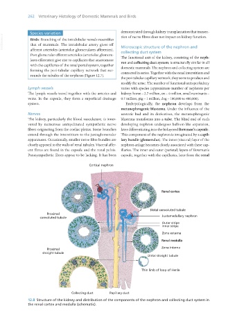
12.8 Structure of the kidney and distribution of the components of the nephron and collecting duct system in
the renal cortex and medulla (schematic).
Vet Histology.indb 262 16/07/2019 15:03