
Page 283 - Veterinary Histology of Domestic Mammals and Birds, 5th Edition
P. 283
Urinary system (organa urinaria) 265
The capillary tuft of the glomerulus (rete capillare
VetBooks.ir glomerulare) develops from the division of the afferent
arteriole into four to eight branches. These branches form
up to 50 anastomosing capillary loops. The structural com-
ponents of the glomerulus (Figure 12.9) comprise:
· endothelial cells of the capillary wall,
· glomerular basement membrane,
· podocytes (= inner visceral layer of Bowman’s cap-
sule) and
· intraglomerular mesangial cells with loose connec-
tive tissue.
ENDOTHELIAL CELLS
The wall of the glomerular capillaries is composed of an
extremely thin endothelium with numerous round pores.
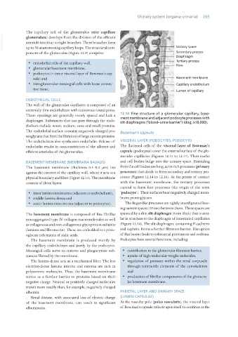
These openings are generally evenly spaced and lack a 12.14 Fine structure of a glomerular capillary, base-
ment membrane and adjacent podocyte processes with
diaphragm. Substances that can pass through the endo- slit diaphragms (‘blood–urine barrier’) (dog; x18,000).
thelium include water, sodium, urea and small proteins.
The endothelial surface contains negatively charged pro- Bowman’s capsule
teoglycans that limit the filtration of large anionic proteins.
The endothelium also synthesises endothelin. Release of VISCERAL LAYER (PODOCYTES, PODOCYTI)
endothelin results in vasoconstriction of the afferent and The flattened cells of the visceral layer of Bowman’s
efferent arterioles of the glomerulus. capsule (podocytes) cover the external surface of the glo-
merular capillaries (Figures 12.15 to 12.17). Their nuclei
BASEMENT MEMBRANE (MEMBRANA BASALIS) and cell bodies bulge into the urinary space. Extending
The basement membrane (thickness 0.1–0.2 μm) lies from the cell bodies are long, actin-rich processes (primary
against the exterior of the capillary wall, where it acts as a processes) that divide to form secondary and tertiary pro-
physical boundary and filter (Figure 12.14). The membrane cesses (Figures 12.14 to 12.16). At the points of contact
consists of three layers: with the basement membrane, the tertiary processes
expand to form foot processes (the origin of the term
· inner lamina rara interna (adjacent to endothelium), ‘podocyte’). Their surfaces bear negatively charged mem-
· middle lamina densa and brane proteoglycans.
· outer lamina rara externa (adjacent to podocytes). The finger-like processes are tightly interdigitated leav-
ing narrow spaces (25 nm) between them. These spaces are
The basement membrane is composed of fine fibrillar spanned by a thin slit diaphragm (6 nm thick) that is simi-
non-aggregated type IV collagen macromolecules as well lar in structure to the diaphragm of fenestrated capillaries
as collagenous and non-collagenous glycoprotein subunits (Figure 12.14). The slit diaphragm, containing P-cadherin
(laminin and fibronectin). These are embedded in a prote- and nephrin, forms a further filtration barrier. Disruption
oglycan-rich matrix of sialic acids. of this barrier leads to substantial proteinuria and oedema.
The basement membrane is produced mainly by Podocytes have several functions, including:
the capillary endothelium and partly by the podocytes.
Mesangial cells serve to remove and phagocytose sub- · contribution to the glomerular filtration barrier,
stances filtered by the membrane. · uptake of high-molecular weight molecules,
The lamina densa acts as a mechanical filter. The less · regulation of pressure within the renal corpuscle
electron-dense lamina interna and externa are rich in through contractile elements of the cytoskeleton
polyanionic molecules. Thus, the basement membrane and
serves as a further barrier to proteins based on their · production of fibrillar components of the glomeru-
negative charge. Neutral or positively charged molecules lar basement membrane.
transit more readily than, for example, negatively charged
albumin. PARIETAL LAYER AND URINARY SPACE
Renal disease, with associated loss of electric charge (LUMEN CAPSULAE)
of the basement membrane, can result in significant At the vascular pole (polus vascularis), the visceral layer
albuminuria. of Bowman’s capsule reflects upon itself to continue as the
Vet Histology.indb 265 16/07/2019 15:03