Page 288 - Veterinary Histology of Domestic Mammals and Birds, 5th Edition
P. 288
270  Veterinary Histology of Domestic Mammals and Birds


       VetBooks.ir


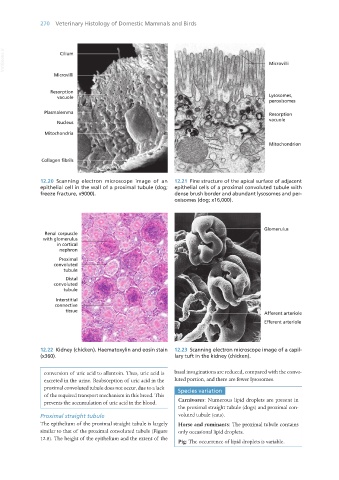
                   12.20  Scanning electron microscope image of an   12.21  Fine structure of the apical surface of adjacent
                   epithelial cell in the wall of a proximal tubule (dog;   epithelial cells of a proximal convoluted tubule with
                   freeze fracture, x9000).                       dense brush border and abundant lysosomes and per-
                                                                  oxisomes (dog; x16,000).


                   12.22  Kidney (chicken). Haematoxylin and eosin stain   12.23  Scanning electron microscope image of a capil-
                   (x360).                                        lary tuft in the kidney (chicken).


                     conversion of uric acid to allantoin. Thus, uric acid is   basal invaginations are reduced, compared with the convo-
                     excreted in the urine. Reabsorption of uric acid in the   luted portion, and there are fewer lysosomes.
                     proximal convoluted tubule does not occur, due to a lack   Species variation
                     of the required transport mechanism in this breed. This
                     prevents the accumulation of uric acid in the blood.  Carnivores: Numerous lipid droplets are present in
                                                                    the proximal straight tubule (dogs) and proximal con-
                   Proximal straight tubule                         voluted tubule (cats).
                   The epithelium of the proximal straight tubule is largely   Horse and ruminants: The proximal tubule contains
                   similar to that of the proximal convoluted tubule (Figure   only occasional lipid droplets.
                   12.8). The height of the epithelium and the extent of the
                                                                    Pig: The occurrence of lipid droplets is variable.


       Vet Histology.indb   270                                                                                  16/07/2019   15:03

