
Page 270 - Veterinary Histology of Domestic Mammals and Birds, 5th Edition
P. 270
252 Veterinary Histology of Domestic Mammals and Birds
Gas exchange interface Alveolar ducts and alveolar sacs
VetBooks.ir Having passed through the conducting airways, inspired The final respiratory bronchioles branch to form up to ten
air reaches the region in which gas exchange takes place. alveolar ducts (ductus alveolares) (Figure 11.22). The walls
The components of this interface (Figures 11.22 to 11.27 of the alveolar ducts consist of closely spaced openings,
and Table 11.2) comprise: each leading into an alveolus lined with simple squamous
epithelium (alveolar epithelium). Externally, the alveolar
· respiratory bronchioles with: ducts are surrounded by a network of collagen fibres with
− simple epithelium, some elastic fibres. Together with smooth muscle cells,
− elastic fibres and smooth muscle cells, this network acts as a sphincter. Terminal branching (3- to
− the first alveoli with alveolar epithelium, 5-fold) of the alveolar ducts gives rise to alveolar sacs (sac-
· alveolar ducts and alveolar sacs with: culi alveolares), the terminal segments of the respiratory
− simple squamous epithelium (alveolar epithelium) system (Figures 11.22 to 11.24).
and
− elastic fibres and smooth muscle cells. Alveoli (alveoli pulmonis)
Alveoli are the site of gas exchange within the respiratory
Respiratory bronchioles system. These are sac-like evaginations located in the final
The terminal bronchioles divide dichotomously into two segments of the bronchial tree – the respiratory bronchioles,
or more respiratory bronchioles, each of which subse- alveolar ducts and alveolar sacs. Alveolar components include:
quently divides into two (generations I–III). The respiratory
bronchioles are largely similar in structure to the terminal · Type I alveolar epithelial cells (Type I pneumocytes),
bronchioles. They are distinguished by deep outpouchings · Type II alveolar epithelial cells (Type II pneumocytes),
(alveoli) lined by a flattened alveolar epithelium. With each · a surface film of phospholipid (surfactant),
subsequent generation (I–III) the number of alveoli increases. · alveolar macrophages and
The respiratory bronchioles thus serve both as conducting · an inter-alveolar septum with macrophages, capil-
airways and as sites of gas exchange (Figure 11.22). lary networks and pores.
Species variation The walls of alveoli (Figures 11.24, 11.26 and 11.27) are
lined by two cell types.
Carnivores: Respiratory bronchioles are extensively
developed. Type I (squamous) alveolar epithelial cells (pneu-
mocytes) (cellulae respiratoriae or squamosae) are
Horse: Occasional respiratory bronchioles are seen. extremely thin, flattened cells. The nucleus bulges slightly
Ruminants and pig: Respiratory bronchioles are rare. into the lumen (Figure 11.27). Type I cells form a contin-
uous layer that lines approximately 95% of the internal
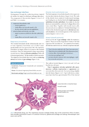
11.21 Bronchiole (lung, ox). The respiratory mucosa is deeply folded, due to the constrictive effect of elastic
fibre bundles and smooth muscle. In contrast to bronchi, glands and cartilage are lacking. Haematoxylin and
eosin stain (x275).
Vet Histology.indb 252 16/07/2019 15:03