Page 268 - Veterinary Histology of Domestic Mammals and Birds, 5th Edition
P. 268
250  Veterinary Histology of Domestic Mammals and Birds


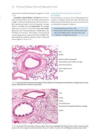
                   contraction of the bronchial muscles (Figures 11.17 and   Bronchioles (bronchioli veri, bronchioli
       VetBooks.ir  11.18).                                       terminales)
                      Incomplete rings of hyaline cartilage form the struc-
                                                                  In the bronchioles, the lumen of the conducting airways is
                   tural core of the bronchi. Near the trachea, they are almost  reduced to a diameter of less than 1 mm. The wall of the
                   complete. With increased branching of the bronchial tree,  small bronchioles (bronchioli veri) and terminal bronchi-
                   the rings become smaller and less substantial (cartilage  oles (bronchioli terminales) comprises:
                   ‘plates’), eventually becoming fragmented. The cartilage
                   fragments are connected by predominantly elastic fibres   ·  ciliated epithelium with Clara cells,
                   that project into the perichondrium, contributing to the   ·  lamina propria with elastic fibres and smooth muscle
                   flexibility of the bronchi. The smallest, most peripheral   cells surrounded by loose connective tissue and
                   cartilage fragments are composed of elastic cartilage. The   ·  no glands or cartilaginous elements.
                   tunica adventitia contains connective tissue, vessels and
                   nerves (Figures 11.16 to 11.19).


                   11.17  Lung (ox). Bronchi are characterised by the presence of mixed glands and hyaline cartilage rings or frag-
                   ments. Haematoxylin and eosin stain (x30).


                   11.18  Lung (calf). The staining technique used in this section highlights the elastic fibres, which are particularly
                   prominent in the larger bronchial branches. Resorcin–fuchsin–nuclear-fast red (Weigert’s elastic) stain (x30).


       Vet Histology.indb   250                                                                                  16/07/2019   15:03

