
Page 389 - Veterinary Histology of Domestic Mammals and Birds, 5th Edition
P. 389
Receptors and sense organs (organa sensuum) 371
VetBooks.ir
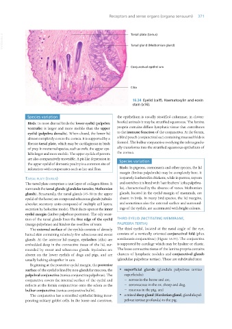
16.34 Eyelid (calf). Haematoxylin and eosin
stain (x16).
Species variation the epithelium is usually stratified columnar; in cloven-
Birds: In most diurnal birds the lower eyelid (palpebra hoofed animals it may be stratified squamous. The lamina
ventralis) is larger and more mobile than the upper propria contains diffuse lymphatic tissue that contributes
eyelid (palpebra dorsalis). When closed, the lower lid to the immune function of the conjunctiva. At the fornix,
almost completely covers the cornea. It is supported by a a blind pouch (conjunctival sac) containing mucosal folds is
fibrous tarsal plate, which may be cartilaginous in birds formed. The bulbar conjunctiva overlying the sclera gradu-
of prey. In nocturnal species, such as owls, the upper eye- ally transforms into the stratified squamous epithelium of
lid is larger and more mobile. The upper eyelids of parrots the cornea.
are also comparatively moveable. A pit-like depression in Species variation
the upper eyelid of domestic poultry is a common site of
infestation with ectoparasites such as lice and fleas. Birds: In pigeons, cormorants and other species, the lid
margin (limbus palpebralis) may be completely bare. It
taRsal Plate (taRsus) is sparsely feathered in chickens, while in parrots, raptors
The tarsal plate comprises a taut layer of collagen fibres. It and ostriches it is lined with ‘hair feathers’ (cilia palpebra-
surrounds the tarsal glands (glandulae tarsales, Meibomian lia), characterised by the absence of vanes. Meibomian
glands). Structurally, the tarsal glands (45–50 in the upper glands, located in the eyelid margin of mammals, are
eyelid of the horse) are compound sebaceous glands (tubulo- absent in birds. In many bird species, the lid margins,
alveolar; secretory units composed of multiple cell layers; and sometimes also the external surface and surround-
secretion by holocrine mode). Their ducts open at the inner ings of the eyelids, are accentuated with bright colours.
eyelid margin (limbus palpebrae posterior). The oily secre-
tion of the tarsal glands lines the free edge of the eyelid THIRD EYELID (NICTITATING MEMBRANE,
(margo palpebrae) and hinders the overflow of tears. PALPEBRA TERTIA)
The external surface of the eyelids consists of densely The third eyelid, located at the nasal angle of the eye,
haired skin containing relatively few sebaceous and sweat consists of a vertically oriented conjunctival fold (plica
glands. At the anterior lid margin, eyelashes (cilia) are semilunaris conjunctivae) (Figure 16.35). The conjunctiva
embedded deep in the connective tissue of the lid, sur- is supported by cartilage which may be hyaline or elastic.
rounded by sweat and sebaceous glands. Eyelashes are The loose connective tissue of the lamina propria contains
absent on the lower eyelids of dogs and pigs, and are clusters of lymphatic nodules and conjunctival glands
usually lacking altogether in cats. (glandulae palpebrae tertiae). These are subdivided into:
Beginning at the posterior eyelid margin, the posterior
surface of the eyelid is lined by non-glandular mucosa, the · superficial glands (glandula palpebrae tertiae
palpebral conjunctiva (tunica conjunctiva palpebrae). The superficialis):
conjunctiva covers the internal surface of the eyelid and − serous in the horse and cat,
reflects at the fornix conjunctivae onto the sclera as the − seromucous in the ox, sheep and dog,
bulbar conjunctiva (tunica conjunctiva bulbi). − mucous in the pig, and
The conjunctiva has a stratified epithelial lining incor- · a mixed deep gland (Harderian gland, glandula pal-
porating solitary goblet cells. In the horse and carnivore, pebrae tertiae profunda) in the pig.
Vet Histology.indb 371 16/07/2019 15:07