
Page 391 - Veterinary Histology of Domestic Mammals and Birds, 5th Edition
P. 391
Receptors and sense organs (organa sensuum) 373
VetBooks.ir · middle ear (auris media):
− tympanic cavity (cavitas tympanica),
− auditory ossicles (ossicula auditus):
− malleus,
− incus,
− stapes,
− auditory tube (tuba auditiva),
· inner ear (auris interna):
− osseous labyrinth (labyrinthus osseus) and
− membranous labyrinth (labyrinthus membranaceus):
− vestibular apparatus – sensory cells for bal-
ance and
− cochlear duct – sensory cells for hearing (Figure
16.38).
External ear (auris externa)
The external ear consists of the pinna, the external acous-
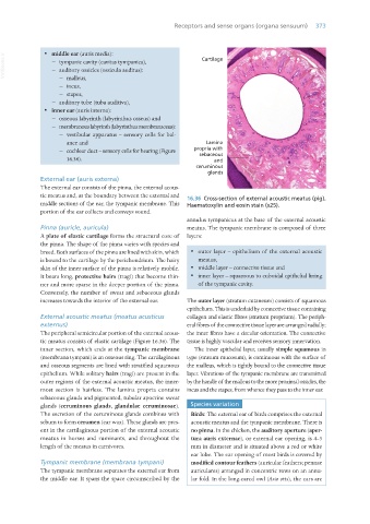
tic meatus and, as the boundary between the external and 16.36 Cross-section of external acoustic meatus (pig).
middle sections of the ear, the tympanic membrane. This Haematoxylin and eosin stain (x25).
portion of the ear collects and conveys sound.
annulus tympanicus at the base of the external acoustic
Pinna (auricle, auricula) meatus. The tympanic membrane is composed of three
A plate of elastic cartilage forms the structural core of layers:
the pinna. The shape of the pinna varies with species and
breed. Both surfaces of the pinna are lined with skin, which · outer layer – epithelium of the external acoustic
is bound to the cartilage by the perichondrium. The hairy meatus,
skin of the inner surface of the pinna is relatively mobile. · middle layer – connective tissue and
It bears long, protective hairs (tragi) that become thin- · inner layer – squamous to cuboidal epithelial lining
ner and more sparse in the deeper portion of the pinna. of the tympanic cavity.
Conversely, the number of sweat and sebaceous glands
increases towards the interior of the external ear. The outer layer (stratum cutaneum) consists of squamous
epithelium. This is underlaid by connective tissue containing
External acoustic meatus (meatus acusticus collagen and elastic fibres (stratum proprium). The periph-
externus) eral fibres of the connective tissue layer are arranged radially;
The peripheral semicircular portion of the external acous- the inner fibres have a circular orientation. The connective
tic meatus consists of elastic cartilage (Figure 16.36). The tissue is highly vascular and receives sensory innervation.
inner section, which ends at the tympanic membrane The inner epithelial layer, usually simple squamous in
(membrana tympani) is an osseous ring. The cartilaginous type (stratum mucosum), is continuous with the surface of
and osseous segments are lined with stratified squamous the malleus, which is tightly bound to the connective tissue
epithelium. While solitary hairs (tragi) are present in the layer. Vibrations of the tympanic membrane are transmitted
outer regions of the external acoustic meatus, the inner- by the handle of the malleus to the more proximal ossicles, the
most section is hairless. The lamina propria contains incus and the stapes, from whence they pass to the inner ear.
sebaceous glands and pigmented, tubular apocrine sweat
glands (ceruminous glands, glandulae ceruminosae). Species variation
The secretion of the ceruminous glands combines with Birds: The external ear of birds comprises the external
sebum to form cerumen (ear wax). These glands are pres- acoustic meatus and the tympanic membrane. There is
ent in the cartilaginous portion of the external acoustic no pinna. In the chicken, the auditory aperture (aper-
meatus in horses and ruminants, and throughout the tura auris externae), or external ear opening, is 4–5
length of the meatus in carnivores. mm in diameter and is situated above a red or white
ear lobe. The ear opening of most birds is covered by
Tympanic membrane (membrana tympani) modified contour feathers (auricular feathers; pennae
The tympanic membrane separates the external ear from auriculares) arranged in concentric rows on an annu-
the middle ear. It spans the space circumscribed by the lar fold. In the long-eared owl (Asio otis), the ears are
Vet Histology.indb 373 16/07/2019 15:07