
Page 392 - Veterinary Histology of Domestic Mammals and Birds, 5th Edition
P. 392
374 Veterinary Histology of Domestic Mammals and Birds
VetBooks.ir covered by a rostral skin fold. This contains striated
muscle that enables the fold to be moved to assist with
localisation of sound. Examination of the auditory
aperture is an important component of ophthalmic
examination in birds.
The external acoustic meatus (meatus acusticus
externus) lies caudal to the quadrate bone. It is approxi-
mately 4–7 mm long.
A small mound in the ventral wall contains an open-
ing for the underlying auricular glands (glandulae
auriculares). The opening of the duct draining these
glands can be visualised under magnification.
The tympanic membrane bulges slightly into the
external ear canal. In the chicken it is 0.012 mm thick
with a surface area of approximately 25 mm . The
2
tympanic membrane is attached to the surrounding
bone and its margins are reinforced by elastic fibres.
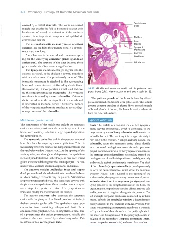
Rostroventrally it incorporates a small, air-filled cav- 16.37 Middle and inner ear in situ within petrous tem-
ity, the sinus pneumaticus marginalis. The tympanic poral bone (pig). Haematoxylin and eosin stain (x16).
membrane is tensed by the m. columellae. This mus-
cle is equivalent to the m. stapedius in mammals and The guttural pouch of the horse is lined by ciliated
is innervated by the facial nerve. The internal surface pseudostratified epithelium with goblet cells. The lamina
of the tympanic membrane is attached to the cartilagi- propria contains bundles of elastic fibres, smooth muscle
nous processes of the columella. cells and glands. A loose, displaceable tunica adventitia
lines the external surface.
Middle ear (auris media) Species variation
The components of the middle ear include the tympanic Birds: The middle ear contains the air-filled tympanic
cavity, the auditory ossicles and the auditory tube. In the cavity (cavitas tympanica), which is connected to the
horse, each auditory tube has a large expanded portion, oropharynx by the auditory tube (tuba auditiva) via the
the guttural pouch. infundibular cleft. The auditory tube is approximately 6
The tympanic cavity lies within the petrous temporal mm long in the chicken. A single auditory ossicle, the
bone. It is lined by simple squamous epithelium. This epi- columella, spans the tympanic cavity. Three flexibly
thelial lining covers the ossicles, the tympanic membrane and interconnected cartilaginous extra-columellar processes
the vestibular window (Figure 16.37). At the opening of the project from the columella to the tympanic membrane as
auditory tube, and throughout this passage, the epithelium the cartilago extracolumellaris. Resembling a tripod, the
is ciliated pseudostratified. In the sheep and carnivore, mixed cartilago extracolumellaris is positioned caudally, ventrally
glands are scattered throughout the lamina propria. The con- and rostrally against the tympanic membrane. The shaft
nective tissue contains abundant capillaries and nerves. of the columella (scapus columellae) expands proximally
The auditory ossicles (malleolus, incus and stapes) to form the basis columellae, which closes the vestibular
develop through endochondral ossification into lamellar bone window (Figure 16.45). Lateral to the opening of the
in which cartilage remnants may be present. Articulations auditory tube, the tympanic cavity houses a small, curved
are present between the bones. The ossicles are covered with vesicular structure, the organum paratympanicum.
simple squamous epithelium. The striated m. tensor tympani Lying parallel to the longitudinal axis of the head, the
and m. stapedius regulate the tension of the tympanic mem- organum paratympanicum contains ciliated sensory cells
brane and modify the movement of the ossicles. and is presumed to register changes in air pressure. The
The narrow auditory tube connects the tympanic left and right tympanic cavities are connected by air-filled
cavity with the pharynx. Its ciliated pseudostratified epi- spaces. In birds, the vestibular window is located imme-
thelium contains goblet cells. The epithelium rests upon diately adjacent to the cochlear window. Pressure from
connective tissue containing collagen and elastic fibres, sound waves striking the tympanic membrane is transmit-
and numerous lymphatic cells. In ungulates, a tubal ton- ted by the base of the columella to the perilymph within
sil is present near the ostium pharyngicum. Initially the the inner ear. Compression of the perilymph results in
auditory tube is surrounded by a short bony collar. This bulging of the secondary tympanic membrane (mem-
transforms into a cartilaginous tube. brana tympanica secundaria) at the cochlear window.
Vet Histology.indb 374 16/07/2019 15:08