
Page 397 - Veterinary Histology of Domestic Mammals and Birds, 5th Edition
P. 397
Receptors and sense organs (organa sensuum) 379
Species variation
VetBooks.ir Birds: As in mammals, the inner ear consists of the
osseous labyrinth and, within it, the membranous
labyrinth. The space between the membranous and
osseous labyrinth is filled with perilymph, while the
membranous labyrinth contains the somewhat viscous
endolymph.
The osseous labyrinth (labyrinthus osseus) is
comprised of the central vestibule (vestibulum), the
ventrally positioned osseous cochlea and the cau-
dodorsally projecting semicircular canals (canales
semicirculares ossei). The avian cochlea is shaped like
a blunt, slightly medially concave and rostrally convex
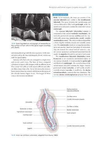
16.44 Scanning electron micrograph of stereocilia on
the surface of hair cells in the spiral organ (courtesy club. The semicircular canals are arranged perpendicu-
of S. Breit). lar to one another. Based on their plane of orientation,
they are termed the rostral vertical (anterior), cau-
dal vertical (posterior) and lateral horizontal (lateral)
and eventually merge with the stria vascularis. At the inter- canals. An ampulla is located at one end of each canal.
nal spiral sulcus, the inner phalangeal cells are continuous The membranous labyrinth (labyrinthus mem-
with the spiral limbus. branaceus) is an extensively partitioned replica of
Sensory cells (hair cells) are arranged in a single inner the osseous labyrinth. It is surrounded by perilymph
and several outer rows. The base of these elongated and filled with endolymph. The central section of the
cylindrical receptor cells synapses with an afferent nerve membranous labyrinth contains the larger, dorsally
fibre (outer hair cells) or with several afferent and effer- positioned tubular utriculus, as well as the smaller,
ent fibres (inner hair cells). Up to 100 stereocilia (sensory ventrally situated sacculus. A small tube, the ductus
hairs) project from the sensory cells between the spaces of utriculosaccularis, connects the two chambers. The
the reticular lamina (Figure 16.44). The longest of these narrow ductus endolymphaticus projects from the
contact the tectorial membrane.
16.45 Inner ear (chicken; schematic; adapted from Evans, 1982).
Vet Histology.indb 379 16/07/2019 15:08