
Page 402 - Veterinary Histology of Domestic Mammals and Birds, 5th Edition
P. 402
384 Veterinary Histology of Domestic Mammals and Birds
VetBooks.ir
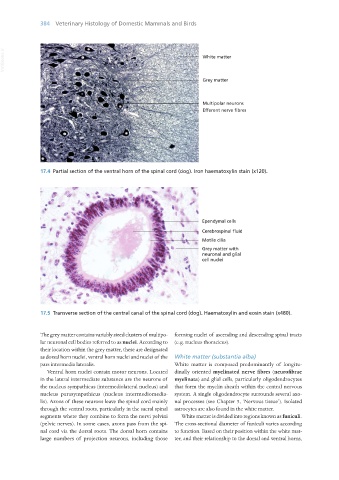
17.4 Partial section of the ventral horn of the spinal cord (dog). Iron haematoxylin stain (x120).
17.5 Transverse section of the central canal of the spinal cord (dog). Haematoxylin and eosin stain (x480).
The grey matter contains variably sized clusters of multipo- forming nuclei of ascending and descending spinal tracts
lar neuronal cell bodies referred to as nuclei. According to (e.g. nucleus thoracicus).
their location within the grey matter, these are designated
as dorsal horn nuclei, ventral horn nuclei and nuclei of the White matter (substantia alba)
pars intermedia lateralis. White matter is composed predominantly of longitu-
Ventral horn nuclei contain motor neurons. Located dinally oriented myelinated nerve fibres (neurofibrae
in the lateral intermediate substance are the neurons of myelinata) and glial cells, particularly oligodendrocytes
the nucleus sympathicus (intermediolateral nucleus) and that form the myelin sheath within the central nervous
nucleus parasympathicus (nucleus intermediomedia- system. A single oligodendrocyte surrounds several axo-
lis). Axons of these neurons leave the spinal cord mainly nal processes (see Chapter 5, ‘Nervous tissue’). Isolated
through the ventral roots, particularly in the sacral spinal astrocytes are also found in the white matter.
segments where they combine to form the nervi pelvini White matter is divided into regions known as funiculi.
(pelvic nerves). In some cases, axons pass from the spi- The cross-sectional diameter of funiculi varies according
nal cord via the dorsal roots. The dorsal horn contains to function. Based on their position within the white mat-
large numbers of projection neurons, including those ter, and their relationship to the dorsal and ventral horns,
Vet Histology.indb 384 16/07/2019 15:08