
Page 406 - Veterinary Histology of Domestic Mammals and Birds, 5th Edition
P. 406
388 Veterinary Histology of Domestic Mammals and Birds
Peripheral nervous system
VetBooks.ir (pars peripherica, systema nervosum
periphericum)
The peripheral nervous system is composed primarily
of nerve fibres and their associated ganglia. Anatomically,
its two components – the somatic (cerebrospinal) and
autonomic (visceral) nervous systems – are closely associ-
ated within nerve fibre bundles. In functional terms, the
somatic nerve fibres are designated as sensory or motor.
Autonomic nerve fibres are classified as sympathetic or
parasympathetic.
With the exception of the cranial nerves, peripheral
nerve fibres have a segmental embryonic precursor and are
connected with the spinal cord via dorsal and ventral roots.
Efferent motor and autonomic fibres leave the spinal cord
through the ventral root to act upon target organs in the
periphery. Peripheral nerve fibres convey nerve impulses to
the central nervous system via afferent (sensory) pathways.
These fibres pass into the dorsal horn of the spinal cord
(see Veterinary Anatomy of Domestic Animals: Textbook and
Colour Atlas).
At specific locations within the peripheral nervous sys-
tem, clusters of nerve cell bodies (perikarya) form localised
thickenings. Outside the central nervous system, such
accumulations of neuronal cell bodies are referred to as
ganglia. Ganglia are surrounded by a connective tissue
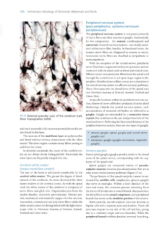
17.11 External granular layer of the cerebrum (cat).
Silver impregnation (x250). capsule that continues as the epi- and perineurium of the
associated nerve. Reflecting the functional divisions of the
peripheral nervous system, there are two types of ganglia:
mid-sized pyramidal cells (neurona pyramidalia media) are
also found in this layer. · sensory ganglia (spinal ganglia and cranial nerve
The neurons of the multiform layer are polymorphic ganglia) and
and blend without obvious demarcation with the white · autonomic ganglia (ganglia autonomica, vegetative
matter. This inner region contains many fibres passing to ganglia).
and from the cortex.
In domestic mammals, the layers of the cerebral cor- Sensory ganglia
tex are not always clearly distinguishable. Particularly the Paired spinal ganglia (ganglia spinalia) reside in the dorsal
inner layers are frequently merged into one. roots of the spinal nerves, corresponding with the seg-
ments of the spinal cord.
Cerebral white matter Spinal ganglia are composed mainly of pseudo-
(corpus medullare cerebri) unipolar neurons (neurona pseudounipolaria) that act as
The size of the brain is influenced considerably by the relay points within sensory pathways (Figure 17.12).
cerebral white matter. The greater the degree of devel- The perikaryon of the pseudo-unipolar neuron is sur-
opment of the cerebrum, the more developed the white rounded by satellite cells (amphicytes, gliocyti ganglii)
matter relative to the cerebral cortex. As with the spinal that serve as neuroglia. Within a short distance of the
cord, the white matter of the cerebrum is composed of neuronal soma, the common process extending from
nerve fibres and glial cells. Oligodendrocytes form the the nerve cell divides into a central branch, that passes into
myelin sheaths; astrocytes (protoplasmic, fibrous) pro- the dorsal horn as the axonal component, and a peripheral
vide mechanical and metabolic support for the neurons. branch that serves as the dendritic portion of the neuron.
Association, commissural and projection fibres within the Embryonically, pseudo-unipolar neurons develop as
white matter cannot be distinguished with the light micro- bipolar cells with a separate axon and dendrite. These cell
scope (refer to Veterinary Anatomy of Domestic Animals: processes migrate to one side of the cell and fuse, giving
Textbook and Colour Atlas). rise to a common origin and two branches. While the
peripheral branch exhibits dendritic terminal branching,
Vet Histology.indb 388 16/07/2019 15:08