
Page 403 - Veterinary Histology of Domestic Mammals and Birds, 5th Edition
P. 403
Nervous system (systema nervosum) 385
these are termed the dorsal funiculus (funiculus dorsalis), Purkinje cells give off an axon into the granule cell layer
VetBooks.ir lateral funiculus (funiculus lateralis) and ventral funiculus and send two or, less frequently, three large dendrites into
(funiculus ventralis). The funiculi contain ascending and the molecular layer. The dendrites arborise into networks
that extend to the surface of the cerebellum. The axons,
descending nerve tracts.
which become myelinated within the Purkinje cell layer,
Cerebellum are the only efferent fibres of the cerebellum and extend
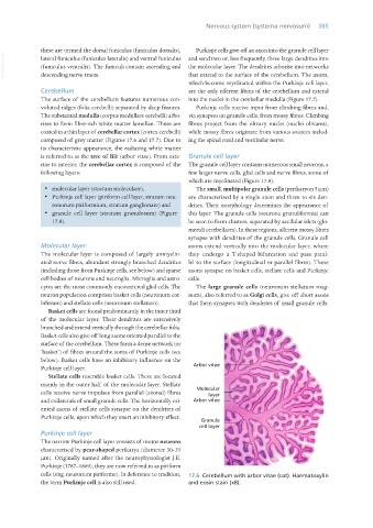
The surface of the cerebellum features numerous con- into the nuclei in the cerebellar medulla (Figure 17.7).
voluted ridges (folia cerebelli) separated by deep fissures. Purkinje cells receive input from climbing fibres and,
The substantial medulla (corpus medullare cerebelli) arbo- via synapses on granule cells, from mossy fibres. Climbing
rises to form fibre-rich white matter lamellae. These are fibres project from the olivary nuclei (nuclei olivares),
coated in a thin layer of cerebellar cortex (cortex cerebelli) while mossy fibres originate from various sources includ-
composed of grey matter (Figures 17.6 and 17.7). Due to ing the spinal cord and vestibular nerve.
its characteristic appearance, the radiating white matter
is referred to as the tree of life (arbor vitae). From exte- Granule cell layer
rior to interior, the cerebellar cortex is composed of the The granule cell layer contains numerous small neurons, a
following layers: few larger nerve cells, glial cells and nerve fibres, some of
which are myelinated (Figure 17.8).
· molecular layer (stratum moleculare), The small, multipolar granule cells (perikaryon 5 μm)
· Purkinje cell layer (piriform cell layer, stratum neu- are characterised by a single axon and three to six den-
ronorum piriformium, stratum ganglionare) and drites. Their morphology determines the appearance of
· granule cell layer (stratum granulosum) (Figure this layer. The granule cells (neurona granuliformia) can
17.8). be seen to form clusters, separated by acellular islets (glo-
meruli cerebellares). In these regions, afferent mossy fibres
synapse with dendrites of the granule cells. Granule cell
Molecular layer axons extend vertically into the molecular layer, where
The molecular layer is composed of largely unmyelin- they undergo a T-shaped bifurcation and pass paral-
ated nerve fibres, abundant strongly branched dendrites lel to the surface (longitudinal or parallel fibres). These
(including those from Purkinje cells, see below) and sparse axons synapse on basket cells, stellate cells and Purkinje
cell bodies of neurons and neuroglia. Microglia and astro- cells.
cytes are the most commonly encountered glial cells. The The large granule cells (neuronum stellatum mag-
neuron population comprises basket cells (neuronum cor- num), also referred to as Golgi cells, give off short axons
biferum) and stellate cells (neuronum stellatum). that form synapses with dendrites of small granule cells.
Basket cells are found predominantly in the inner third
of the molecular layer. Their dendrites are extensively
branched and extend vertically through the cerebellar folia.
Basket cells also give off long axons oriented parallel to the
surface of the cerebellum. These form a dense network (or
‘basket’) of fibres around the soma of Purkinje cells (see
below). Basket cells have an inhibitory influence on the
Purkinje cell layer.
Stellate cells resemble basket cells. These are located
mainly in the outer half of the molecular layer. Stellate
cells receive nerve impulses from parallel (axonal) fibres
and collaterals of small granule cells. The horizontally ori-
ented axons of stellate cells synapse on the dendrites of
Purkinje cells, upon which they exert an inhibitory effect.
Purkinje cell layer
The narrow Purkinje cell layer consists of motor neurons
characterised by pear-shaped perikarya (diameter 30–35
μm). Originally named after the neurophysiologist J.E.
Purkinje (1787–1869), they are now referred to as piriform
cells (sing. neuronum piriforme). In deference to tradition, 17.6 Cerebellum with arbor vitae (cat). Haematoxylin
the term Purkinje cell is also still used. and eosin stain (x8).
Vet Histology.indb 385 16/07/2019 15:08