
Page 405 - Veterinary Histology of Domestic Mammals and Birds, 5th Edition
P. 405
Nervous system (systema nervosum) 387
VetBooks.ir
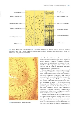
17.9 Layers of the cerebrum (schematic). A: Golgi silver impregnation method showing branching of nerve
processes, B: Nissl stain showing neuronal endoplasmic reticulum, C: staining of myelin sheath to illustrate the
architecture of myelinated nerve fibres.
surface. Together with the terminal branches of associa-
tion and commissural fibres, these give rise to a tangentially
oriented meshwork. The nerve cells act as interneurons.
Also present in the molecular layer are dendrites of pyram-
idal cells located in deeper layers of the cortex. Fibrous
astrocytes form the superficial glial limiting membrane
(membrana limitans gliae superficialis).
The external granular layer consists of numerous small
(10–12 μm) pyramidal nerve cells (neurona pyramidalia
parva). The processes of these cells pass into the superficial
and deeper cortical layers. In addition to small collateral
branches, the cells have a principal dendrite that gives rise
to the pyramidal shape of the cell. The external pyramidal
layer contains mid-sized and large pyramidal cells (neu-
rona pyramidalia), the size of the cell increasing towards
the inner portion of the layer (20–40 μm). The external
pyramidal layer is generally the thickest layer of the cer-
ebral cortex. The internal granular layer is composed of
small neurons of variable morphology. These cells serve
as interneurons, their processes forming macroscopically
visible stria that run parallel to the surface.
The internal pyramidal layer is characterised by large
pyramidal neurons (neurona pyramidalia magna, 80–120
μm). The axons given off by these cells form a component
of the pyramidal tracts. Structurally, the pyramidal neu-
rons of the internal pyramidal layer resemble those of the
17.10 Cerebrum (dog). Golgi stain (x10).
ventral horn of the spinal cord. Regional accumulations of
Vet Histology.indb 387 16/07/2019 15:08