
Page 43 - Equine Clinical Medicine, Surgery and Reproduction, 2nd Edition
P. 43
18 CHAPTER 1
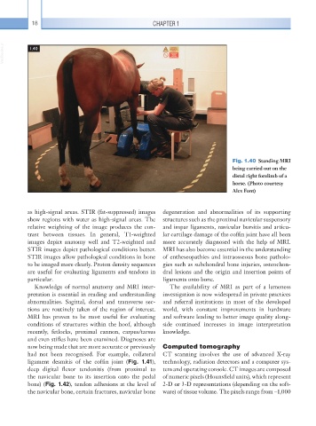
VetBooks.ir 1.40
Fig. 1.40 Standing MRI
being carried out on the
distal right forelimb of a
horse. (Photo courtesy
Alex Font)
as high-signal areas. STIR (fat-suppressed) images degeneration and abnormalities of its supporting
show regions with water as high-signal areas. The structures such as the proximal navicular suspensory
relative weighting of the image produces the con- and impar ligaments, navicular bursitis and articu-
trast between tissues. In general, T1-weighted lar cartilage damage of the coffin joint have all been
images depict anatomy well and T2-weighted and more accurately diagnosed with the help of MRI.
STIR images depict pathological conditions better. MRI has also become essential in the understanding
STIR images allow pathological conditions in bone of entheseopathies and intraosseous bone patholo-
to be imaged more clearly. Proton density sequences gies such as subchondral bone injuries, osteochon-
are useful for evaluating ligaments and tendons in dral lesions and the origin and insertion points of
particular. ligaments onto bone.
Knowledge of normal anatomy and MRI inter- The availability of MRI as part of a lameness
pretation is essential in reading and understanding investigation is now widespread in private practices
abnormalities. Sagittal, dorsal and transverse sec- and referral institutions in most of the developed
tions are routinely taken of the region of interest. world, with constant improvements in hardware
MRI has proven to be most useful for evaluating and software leading to better image quality along-
conditions of structures within the hoof, although side continued increases in image interpretation
recently, fetlocks, proximal cannon, carpus/tarsus knowledge.
and even stifles have been examined. Diagnoses are
now being made that are more accurate or previously Computed tomography
had not been recognised. For example, collateral CT scanning involves the use of advanced X-ray
ligament desmitis of the coffin joint (Fig. 1.41), technology, radiation detectors and a computer sys-
deep digital flexor tendonitis (from proximal to tem and operating console. CT images are composed
the navicular bone to its insertion onto the pedal of numeric pixels (Hounsfield units), which represent
bone) (Fig. 1.42), tendon adhesions at the level of 2-D or 3-D representations (depending on the soft-
the navicular bone, certain fractures, navicular bone ware) of tissue volume. The pixels range from –1,000