
Page 42 - Equine Clinical Medicine, Surgery and Reproduction, 2nd Edition
P. 42
Musculoskeletal system: 1.1 A pproach to the lame horse 17
VetBooks.ir 1.38 1.39
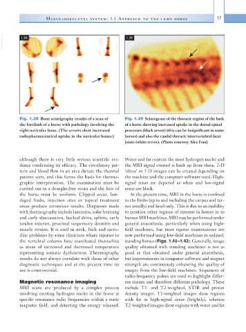
Fig. 1.38 Bone scintigraphy results of a scan of Fig. 1.39 Scintogram of the thoracic region of the back
the forelimb of a horse with pathology involving the of a horse showing increased uptake in the dorsal spinal
right navicular bone. (The arrows show increased processes (black arrow) (this can be insignificant in some
radiopharmaceutical uptake in the navicular bones.) horses) and also the caudal thoracic intervertebral facet
joints (white arrow). (Photo courtesy Alex Font)
although there is very little serious scientific evi- Water and fat contain the most hydrogen nuclei and
dence confirming its efficacy. The circulatory pat- the MRI signal created is built up from these. 2-D
tern and blood flow in an area dictate the thermal ‘slices’ or 3-D images can be created depending on
pattern seen, and this forms the basis for thermo- the machine and the computer software used. High-
graphic interpretation. The examination must be signal areas are depicted as white and low- signal
carried out in a draught-free room and the hair of areas are black.
the horse must be uniform. Clipped areas, ban- At the present time, MRI in the horse is confined
daged limbs, injection sites or topical treatment to the limbs (up to and including the carpus and tar-
areas produce erroneous results. Diagnoses made sus usually) and head only. This is due to an inability
with thermography include laminitis, solar bruising to position other regions of interest in horses in to
and early abscessation, bucked shins, splints, early human MRI machines. MRI may be performed under
tendon injuries, proximal suspensory desmitis and general anaesthesia, particularly when using high-
muscle strains. It is used in neck, back and sacro- field machines, but most equine examinations are
iliac problems by some clinicians where injuries to now performed using low-field machines in sedated,
the vertebral column have manifested themselves standing horses (Figs. 1.40–1.42). Generally, image
as areas of increased and decreased temperature quality obtained with standing machines is not as
representing somatic dysfunction. Thermography good as that obtained under general anaesthesia,
results do not always correlate with those of other but improvements in computer software and magnet
diagnostic techniques and at the present time its strength are continuously enhancing the quality of
use is controversial. images from the low-field machines. Sequences of
radio-frequency pulses are used to highlight differ-
Magnetic resonance imaging ent tissues and therefore different pathology. These
MRI scans are produced by a complex process include T1- and T2-weighted, STIR and proton
involving exciting hydrogen nuclei in the horse at density images. T1-weighted images show regions
specific resonance radio frequencies within a static with fat as high-signal areas (brightly), whereas
magnetic field, and detecting the energy released. T2-weighted images show regions with water and fat