
Page 40 - Equine Clinical Medicine, Surgery and Reproduction, 2nd Edition
P. 40
Musculoskeletal system: 1.1 A pproach to the lame horse 15
VetBooks.ir are now commonly available for most machines and are image soft-tissue structures (e.g. tendons, liga-
Ultrasonography is most commonly used to
sufficient for most examinations. The term hypoechoic
denotes a decreased echogenicity of the tissue (darker
linings [Figs. 1.34, 1.35], localised soft-tissue swell-
image), anechoic denotes no echogenicity (i.e. fluid ments [Figs. 1.32, 1.33], joint capsules, synovial
[black image]) and hyperechoic denotes an increased ings [Fig. 1.36], muscles, nerves and blood vessels).
echogenicity (brighter image). Transverse and longitudinal images are essential in
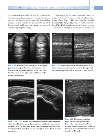
1.32 1.33
Fig. 1.32 Transverse ultrasonogram of the upper Fig. 1.33 Sagittal longitudinal ultrasonogram of the
palmar metacarpus of a racehorse with acute-onset same horse demonstrating the lesion in the right limb
lameness and soft-tissue swelling of the area. Note and confirming the length of ligament that is injured.
the core lesion in the right upper mid-body of the
suspensory ligament.
1.34 1.35 1.36
Fig. 1.36 Ultrasonogram of an
Figs. 1.34, 1.35 Sagittal (1.34) and oblique (1.35) ultrasonograms injection abscess in the neck of a
of the dorsal fetlock region of a young Thoroughbred with sepsis of horse following vaccination. Note
the joint. Note the distended dorsal metacarpophalangeal joint filled the oval shaped hypoechogenic abscess
with hyperechoic joint fluid and the hyperplasia of the dorsal synovial (between the crosses) deep within the
membrane. muscles of the neck.