
Page 38 - Equine Clinical Medicine, Surgery and Reproduction, 2nd Edition
P. 38
Musculoskeletal system: 1.1 A pproach to the lame horse 13
VetBooks.ir 1.26 1.27
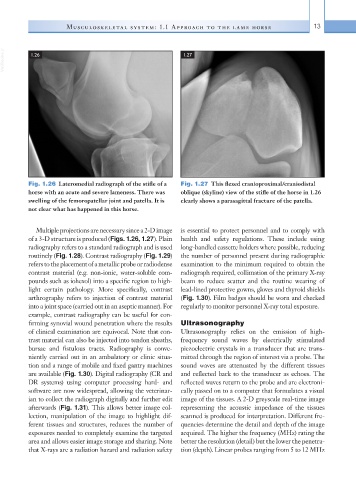
Fig. 1.26 Lateromedial radiograph of the stifle of a Fig. 1.27 This flexed cranioproximal/craniodistal
horse with an acute and severe lameness. There was oblique (skyline) view of the stifle of the horse in 1.26
swelling of the femoropatellar joint and patella. It is clearly shows a parasagittal fracture of the patella.
not clear what has happened in this horse.
Multiple projections are necessary since a 2-D image is essential to protect personnel and to comply with
of a 3-D structure is produced (Figs. 1.26, 1.27). Plain health and safety regulations. These include using
radiography refers to a standard radiograph and is used long-handled cassette holders where possible, reducing
routinely (Fig. 1.28). Contrast radiography (Fig. 1.29) the number of personnel present during radiographic
refers to the placement of a metallic probe or radiodense examination to the minimum required to obtain the
contrast material (e.g. non-ionic, water-soluble com- radiograph required, collimation of the primary X-ray
pounds such as iohexol) into a specific region to high- beam to reduce scatter and the routine wearing of
light certain pathology. More specifically, contrast lead-lined protective gowns, gloves and thyroid shields
arthrography refers to injection of contrast material (Fig. 1.30). Film badges should be worn and checked
into a joint space (carried out in an aseptic manner). For regularly to monitor personnel X-ray total exposure.
example, contrast radiography can be useful for con-
firming synovial wound penetration where the results Ultrasonography
of clinical examination are equivocal. Note that con- Ultrasonography relies on the emission of high-
trast material can also be injected into tendon sheaths, frequency sound waves by electrically stimulated
bursae and fistulous tracts. Radiography is conve- piezoelectric crystals in a transducer that are trans-
niently carried out in an ambulatory or clinic situa- mitted through the region of interest via a probe. The
tion and a range of mobile and fixed gantry machines sound waves are attenuated by the different tissues
are available (Fig. 1.30). Digital radiography (CR and and reflected back to the transducer as echoes. The
DR systems) using computer processing hard- and reflected waves return to the probe and are electroni-
software are now widespread, allowing the veterinar- cally passed on to a computer that formulates a visual
ian to collect the radiograph digitally and further edit image of the tissues. A 2-D greyscale real-time image
afterwards (Fig. 1.31). This allows better image col- representing the acoustic impedance of the tissues
lection, manipulation of the image to highlight dif- scanned is produced for interpretation. Different fre-
ferent tissues and structures, reduces the number of quencies determine the detail and depth of the image
exposures needed to completely examine the targeted acquired. The higher the frequency (MHz) rating the
area and allows easier image storage and sharing. Note better the resolution (detail) but the lower the penetra-
that X-rays are a radiation hazard and radiation safety tion (depth). Linear probes ranging from 5 to 12 MHz