
Page 1172 - Clinical Small Animal Internal Medicine
P. 1172
1110 Section 10 Renal and Genitourinary Disease
Pomeranian, shih tzu, etc.) at risk for calcium oxalate Point‐of‐care diagnostics are especially important in
VetBooks.ir stones. Because of their strong association with urinary the initial stages of assessing the sick obstructed patient.
This should include packed cell volume and total pro
tract infection (UTI), female dogs are much more likely
to have struvite urolithiasis. Dalmatians and breeds pre
status, and/or kidney values (if available). Expected
disposed to portosystemic shunts (e.g., Yorkshire terri tein, as well as assessment of electrolyte, acid–base
ers) should be considered for the potential of urate stones. abnormalities include azotemia, hyperkalemia, and met
Lower urinary tract neoplasia is reported to have a higher abolic acidosis. An ECG should be placed to evaluate the
prevalence in Scottish terriers, Shetland sheepdogs, wire‐ effects of hyperkalemia on electrical conduction in the
haired fox terriers, and West Highland white terrier. heart, even if the patient is not demonstrating bradycar
dia. Progressive changes that can be seen associated with
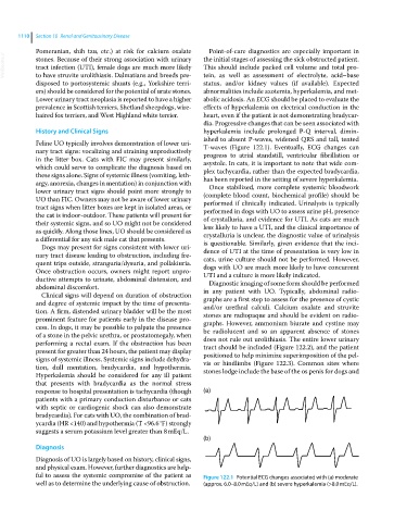
History and Clinical Signs hyperkalemia include prolonged P‐Q interval, dimin
ished to absent P‐waves, widened QRS and tall, tented
Feline UO typically involves demonstration of lower uri T‐waves (Figure 122.1). Eventually, ECG changes can
nary tract signs: vocalizing and straining unproductively progress to atrial standstill, ventricular fibrillation or
in the litter box. Cats with FIC may present similarly, asystole. In cats, it is important to note that wide com
which could serve to complicate the diagnosis based on plex tachycardia, rather than the expected bradycardia,
these signs alone. Signs of systemic illness (vomiting, leth has been reported in the setting of severe hyperkalemia.
argy, anorexia, changes in mentation) in conjunction with Once stabilized, more complete systemic bloodwork
lower urinary tract signs should point more strongly to (complete blood count, biochemical profile) should be
UO than FIC. Owners may not be aware of lower urinary performed if clinically indicated. Urinalysis is typically
tract signs when litter boxes are kept in isolated areas, or performed in dogs with UO to assess urine pH, presence
the cat is indoor‐outdoor. These patients will present for of crystalluria, and evidence for UTI. As cats are much
their systemic signs, and so UO might not be considered less likely to have a UTI, and the clinical importance of
as quickly. Along those lines, UO should be considered as crystalluria is unclear, the diagnostic value of urinalysis
a differential for any sick male cat that presents. is questionable. Similarly, given evidence that the inci
Dogs may present for signs consistent with lower uri
nary tract disease leading to obstruction, including fre dence of UTI at the time of presentation is very low in
cats, urine culture should not be performed. However,
quent trips outside, stranguria/dysuria, and pollakiuria. dogs with UO are much more likely to have concurrent
Once obstruction occurs, owners might report unpro UTI and a culture is more likely indicated.
ductive attempts to urinate, abdominal distension, and Diagnostic imaging of some form should be performed
abdominal discomfort. in any patient with UO. Typically, abdominal radio
Clinical signs will depend on duration of obstruction
and degree of systemic impact by the time of presenta graphs are a first step to assess for the presence of cystic
and/or urethral calculi. Calcium oxalate and struvite
tion. A firm, distended urinary bladder will be the most stones are radiopaque and should be evident on radio
prominent feature for patients early in the disease pro graphs. However, ammonium biurate and cystine may
cess. In dogs, it may be possible to palpate the presence be radiolucent and so an apparent absence of stones
of a stone in the pelvic urethra, or prostatomegaly, when does not rule out urolithiasis. The entire lower urinary
performing a rectal exam. If the obstruction has been tract should be included (Figure 122.2), and the patient
present for greater than 24 hours, the patient may display positioned to help minimize superimposition of the pel
signs of systemic illness. Systemic signs include dehydra vis or hindlimbs (Figure 122.3). Common sites where
tion, dull mentation, bradycardia, and hypothermia. stones lodge include the base of the os penis for dogs and
Hyperkalemia should be considered for any ill patient
that presents with bradycardia as the normal stress
response to hospital presentation is tachycardia (though (a)
patients with a primary conduction disturbance or cats
with septic or cardiogenic shock can also demonstrate
bradycardia). For cats with UO, the combination of brad
ycardia (HR <140) and hypothermia (T <96.6 °F) strongly
suggests a serum potassium level greater than 8 mEq/L.
(b)
Diagnosis
Diagnosis of UO is largely based on history, clinical signs,
and physical exam. However, further diagnostics are help
ful to assess the systemic compromise of the patient as Figure 122.1 Potential ECG changes associated with (a) moderate
well as to determine the underlying cause of obstruction. (approx. 6.0–8.0 mEq/L) and (b) severe hyperkalemia (>8.0 mEq/L).