
Page 1173 - Clinical Small Animal Internal Medicine
P. 1173
122 Obstructive Uropathy 1111
(a) (b)
VetBooks.ir
Figure 122.2 Lateral abdominal radiographic projection of a cat depicting (a) normal abdominal positioning technique and (b)
positioning to include the entire lower urinary tract. Note the urolith evident in the distal urethra (arrow).
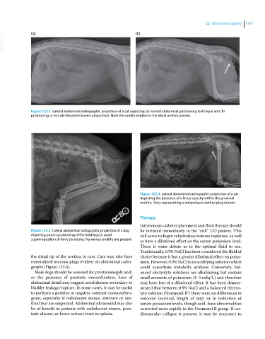
Figure 122.4 Lateral abdominal radiographic projection of a cat
depicting the presence of a linear opacity within the proximal
urethra, likely representing a mineralized urethral plug (arrow).
Therapy
Intravenous catheter placement and fluid therapy should
Figure 122.3 Lateral abdominal radiographic projection of a dog be initiated immediately in the “sick” UO patient. This
depicting proper positioning of the hind legs to avoid will serve to begin rehydration/volume repletion, as well
superimposition of bony structures. Numerous uroliths are present. as have a dilutional effect on the serum potassium level.
There is some debate as to the optimal fluid to use.
Traditionally, 0.9% NaCl has been considered the fluid of
the distal tip of the urethra in cats. Cats may also have choice because it has a greater dilutional effect on potas
mineralized mucous plugs evident on abdominal radio sium. However, 0.9% NaCl is an acidifying solution which
graphs (Figure 122.4). could exacerbate metabolic acidosis. Conversely, bal
Male dogs should be assessed for prostatomegaly and/ anced electrolyte solutions are alkalinizing but contain
or the presence of prostatic mineralization. Loss of small amounts of potassium (4–5 mEq/L) and therefore
abdominal detail may suggest uroabdomen secondary to may have less of a dilutional effect. It has been demon
bladder leakage/rupture. In some cases, it may be useful strated that between 0.9% NaCl and a balanced electro
to perform a positive or negative contrast cystourethro lyte solution (Normosol R®) there were no differences in
gram, especially if radiolucent stones, stricture or ure outcome (survival, length of stay) or in reduction of
thral tear are suspected. Abdominal ultrasound may also serum potassium levels, though acid–base abnormalities
be of benefit in patients with radiolucent stones, pros corrected more rapidly in the Normosol R group. If car
tatic disease, or lower urinary tract neoplasia. diovascular collapse is present, it may be necessary to