
Page 1175 - Clinical Small Animal Internal Medicine
P. 1175
122 Obstructive Uropathy 1113
polytetrafluoroethylene) can be used to initially relieve For catheterization and deobstruction in dogs, general
VetBooks.ir the obstruction. Given the very rigid nature of polypro anesthesia can be especially important to ensure optimal
urethral relaxation. The prepuce or vulva should be
pylene catheters, care should be taken not to use too
much force in advancing. For comfort, as well as associ
red rubber or Foley catheter is typically used, though in
ated inflammation and irritation, these catheters should cleaned and flushed to decrease risk of contamination. A
also not be left in place. With regard to optimal catheter some circumstances a more rigid catheter may be needed
size, there is some evidence to suggest that use of a 3.5 Fr (with potential increased risk of urethral trauma). Similar
urinary catheter may be associated with less risk of to the process described for cats, hydropulsion with
immediate reobstruction compared to 5 Fr. However, lubricated saline should be the primary means of retro
another study failed to show this association. pulsing any physical obstruction. In male dogs, it is help
When passing the catheter, aggressive flushing (rather ful to pinch the urethral orifice to prevent antegrade
than force) should be utilized to dilate the urethra and movement of flush solution. An additional useful trick
retropulse/break down any physical component of the (in either male or female dogs) is to have an assistant rec
obstruction. For the flush solution, adding sterile lubri tally apply pressure to the pelvic urethral during hydro
cant to sterile saline and mixing across a three‐way stop pulsion. This allows a build‐up of pressure which, when
cock (in a ratio of 10:1) may help to decrease urethral released, can help dislodge a luminal obstruction.
injury by allowing lubricant to be deposited throughout If urethral catheterization fails due to urethral trauma,
catheter placement (rather than just lubricating the end rupture, or persistent partial obstruction, antegrade ure
of the catheter). Another helpful technique is to pull the thral access can provide a means to deobstruct the
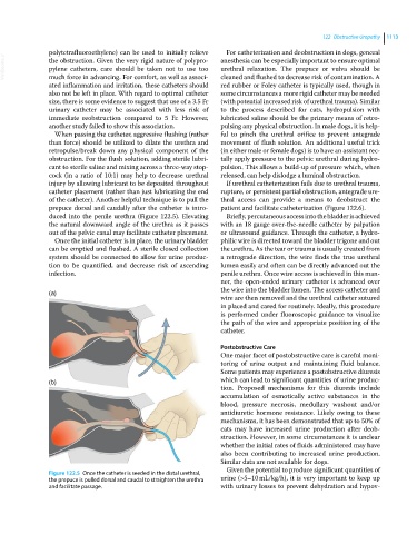
prepuce dorsal and caudally after the catheter is intro patient and facilitate catheterization (Figure 122.6).
duced into the penile urethra (Figure 122.5). Elevating Briefly, percutaneous access into the bladder is achieved
the natural downward angle of the urethra as it passes with an 18 gauge over‐the‐needle catheter by palpation
out of the pelvic canal may facilitate catheter placement. or ultrasound guidance. Through the catheter, a hydro
Once the initial catheter is in place, the urinary bladder philic wire is directed toward the bladder trigone and out
can be emptied and flushed. A sterile closed collection the urethra. As the tear or trauma is usually created from
system should be connected to allow for urine produc a retrograde direction, the wire finds the true urethral
tion to be quantified, and decrease risk of ascending lumen easily and often can be directly advanced out the
infection. penile urethra. Once wire access is achieved in this man
ner, the open‐ended urinary catheter is advanced over
the wire into the bladder lumen. The access catheter and
(a)
wire are then removed and the urethral catheter sutured
in placed and cared for routinely. Ideally, this procedure
is performed under fluoroscopic guidance to visualize
the path of the wire and appropriate positioning of the
catheter.
Postobstructive Care
One major facet of postobstructive care is careful moni
toring of urine output and maintaining fluid balance.
Some patients may experience a postobstructive diuresis
(b) which can lead to significant quantities of urine produc
tion. Proposed mechanisms for this diuresis include
accumulation of osmotically active substances in the
blood, pressure necrosis, medullary washout and/or
antidiuretic hormone resistance. Likely owing to these
mechanisms, it has been demonstrated that up to 50% of
cats may have increased urine production after deob
struction. However, in some circumstances it is unclear
whether the initial rates of fluids administered may have
also been contributing to increased urine production.
Similar data are not available for dogs.
Given the potential to produce significant quantities of
Figure 122.5 Once the catheter is seeded in the distal urethral,
the prepuce is pulled dorsal and caudal to straighten the urethra urine (>5–10 mL/kg/h), it is very important to keep up
and facilitate passage. with urinary losses to prevent dehydration and hypov