
Page 625 - Clinical Small Animal Internal Medicine
P. 625
55 Pancreatitis in the Dog 593
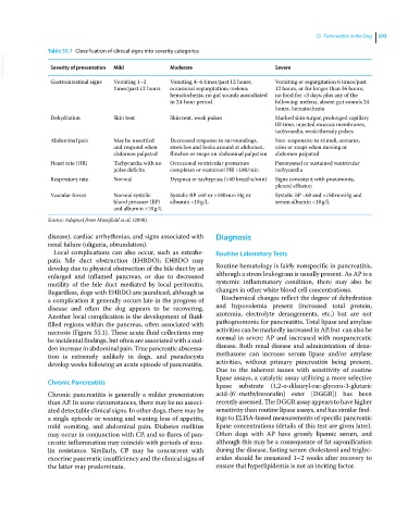
Table 55.1 Classification of clinical signs into severity categories
VetBooks.ir Severity of presentation Mild Moderate Severe
Gastrointestinal signs Vomiting 1–2 Vomiting 4–6 times/past 12 hours; Vomiting or regurgitation 6 times/past
times/past 12 hours occasional regurgitation; melena; 12 hours, or for longer than 36 hours;
hematochezia; no gut sounds auscultated no food for >3 days; plus any of the
in 24‐hour period following: melena, absent gut sounds 24
hours, hematochezia
Dehydration Skin tent Skin tent, weak pulses Marked skin turgor, prolonged capillary
fill time, injected mucous membranes,
tachycardia, weak/thready pulses
Abdominal pain May be unsettled Decreased response to surroundings, Non-responsive to stimuli, screams,
and respond when stretches and looks around at abdomen, cries or snaps when moving or
abdomen palpated flinches or snaps on abdominal palpation abdomen palpated
Heart rate (HR) Tachycardia with no Occasional ventricular premature Paroxysmal or sustained ventricular
pulse deficits complexes or sustained HR >180/min tachycardia
Respiratory rate Normal Dyspnea or tachypnea (>40 breaths/min) Signs consistent with pneumonia,
pleural effusion
Vascular forces Normal systolic Systolic BP <60 or >180 mm Hg or Systolic BP <60 and >180 mmHg and
blood pressure (BP) albumin <18 g/L serum albumin <18 g/L
and albumin >18 g/L
Source: Adapted from Mansfield et al. (2008).
disease), cardiac arrhythmias, and signs associated with Diagnosis
renal failure (oliguria, obtundation).
Local complications can also occur, such as extrahe- Routine Laboratory Tests
patic bile duct obstruction (EHBDO); EHBDO may
develop due to physical obstruction of the bile duct by an Routine hematology is fairly nonspecific in pancreatitis,
enlarged and inflamed pancreas, or due to decreased although a stress leukogram is usually present. As AP is a
motility of the bile duct mediated by local peritonitis. systemic inflammatory condition, there may also be
Regardless, dogs with EHBDO are jaundiced, although as changes in other white blood cell concentrations.
a complication it generally occurs late in the progress of Biochemical changes reflect the degree of dehydration
disease and often the dog appears to be recovering. and hypovolemia present (increased total protein,
Another local complication is the development of fluid‐ azotemia, electrolyte derangements, etc.) but are not
filled regions within the pancreas, often associated with pathognomonic for pancreatitis. Total lipase and amylase
necrosis (Figure 55.1). These acute fluid collections may activities can be markedly increased in AP, but can also be
be incidental findings, but often are associated with a sud- normal in severe AP and increased with nonpancreatic
den increase in abdominal pain. True pancreatic abscessa- disease. Both renal disease and administration of dexa-
tion is extremely unlikely in dogs, and pseudocysts methasone can increase serum lipase and/or amylase
develop weeks following an acute episode of pancreatitis. activities, without primary pancreatitis being present.
Due to the inherent issues with sensitivity of routine
lipase assays, a catalytic assay utilizing a more selective
Chronic Pancreatitis lipase substrate (1,2‐o‐dilauryl‐rac‐glycero‐3‐glutaric
Chronic pancreatitis is generally a milder presentation acid‐{6′‐methylresorufin} ester [DGGR]) has been
than AP. In some circumstances, there may be no associ- recently assessed. The DGGR assay appears to have higher
ated detectable clinical signs. In other dogs, there may be sensitivity than routine lipase assays, and has similar find-
a single episode or waxing and waning loss of appetite, ings to ELISA‐based measurements of specific pancreatic
mild vomiting, and abdominal pain. Diabetes mellitus lipase concentrations (details of this test are given later).
may occur in conjunction with CP, and so flares of pan- Often dogs with AP have grossly lipemic serum, and
creatic inflammation may coincide with periods of insu- although this may be a consequence of fat saponification
lin resistance. Similarly, CP may be concurrent with during the disease, fasting serum cholesterol and triglyc-
exocrine pancreatic insufficiency and the clinical signs of erides should be measured 1–2 weeks after recovery to
the latter may predominate. ensure that hyperlipidemia is not an inciting factor.