
Page 628 - Clinical Small Animal Internal Medicine
P. 628
596 Section 6 Gastrointestinal Disease
(a)
VetBooks.ir
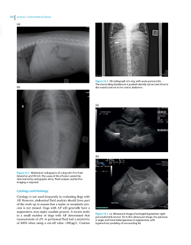
Figure 55.3 VD radiograph of a dog with acute pancreatitis.
The descending duodenum is pushed laterally (arrow) and there is
(b) decreased contrast in the cranial abdomen.
(a)
(b)
Figure 55.2 Abdominal radiographs of a dog with free fluid
(lateral (a) and VD (b)). The cause of the effusion cannot be
determined by radiographs alone. Fluid analysis and further
imaging is required.
Cytology and Histology
Cytology is not used frequently in evaluating dogs with
AP. However, abdominal fluid analysis should form part
of the work‐up to ensure that a septic or neoplastic pro-
cess is not missed. Dogs with AP will generally have a
suppurative, non-septic exudate present. A recent study
in a small number of dogs with AP determined that Figure 55.4 (a) Ultrasound image of enlarged hypoechoic right
pancreatic limb (arrow). (b) In this ultrasound image, the pancreas
measurement of cPL in peritoneal fluid had a sensitivity is larger and more heterogeneous in appearance, with
of 100% when using a cut‐off value >500 μg/L. Caution hyperechoic speckling of surrounding fat.