
Page 348 - Feline diagnostic imaging
P. 348
356 22 Gastrointestinal Disease
that obstruction is present. On ultrasonography, retention fluid opacity may be seen in the caudodorsal thorax. On
of fluid in the stomach and reduced motility can be seen the ventrodorsal projection, there may be a fluid opacity
with obstruction. In one case of feline gastrointestinal located centrally cranial to the diaphragm. Rugal folds
eosinophilic sclerosing fibroplasia (FGESF), the pylorus might be apparent if there is gas within the lumen. Barium
and pyloric antrum had loss of wall layering and the nor- can be given to outline and confirm the presence of a dis-
mal hypoechoic triangular appearance of the pylorus was placed stomach.
not visible. Because this condition is not uncommon, ultra-
sound examinations should include visualization of the 22.3.6 Hiatal Hernia
pylorus [19].
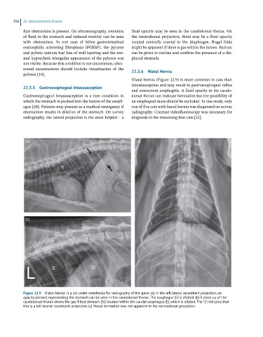
Hiatal hernia (Figure 22.9) is more common in cats than
intussusception and may result in gastroesophageal reflux
22.3.5 Gastroesophageal Intussusception
and concurrent esophagitis. A fluid opacity in the caudo-
Gastroesophageal intussusception is a rare condition in dorsal thorax can indicate herniation but the possibility of
which the stomach is pushed into the lumen of the esoph- an esophageal mass should be excluded. In one study, only
agus [20]. Patients may present as a medical emergency if one of five cats with hiatal hernia was diagnosed on survey
obstruction results in dilation of the stomach. On survey radiography. Contrast videofluoroscopy was necessary for
radiography, the lateral projection is the most helpful – a diagnosis in the remaining four cats [21].
(a) (c)
(b)
Figure 22.9 Hiatal hernia in a cat under anesthesia for radiography of the spine. (a) In the left lateral recumbent projection, an
opacity (arrows) representing the stomach can be seen in the caudodorsal thorax. The esophagus (E) is dilated. (b) A close-up of the
caudodorsal thorax shows the gas-filled stomach (St) located within the caudal esophagus (E), which is dilated. The “L” indicates that
this is a left lateral recumbent projection. (c) Hiatal herniation was not apparent in the ventrodorsal projection.