
Page 351 - Feline diagnostic imaging
P. 351
22.4 Small ntestinal Disorders 359
(a) (b)
(c)
(d)
(e)
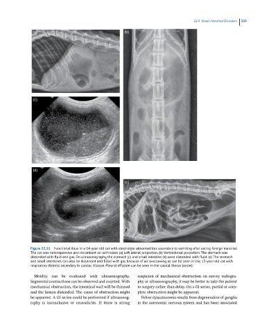
Figure 22.11 Functional ileus in a 14-year-old cat with electrolyte abnormalities secondary to vomiting after eating foreign material.
The cat was nonresponsive and recumbent on admission. (a) Left lateral projection. (b) Ventrodorsal projection. The stomach was
distended with fluid and gas. On ultrasonography, the stomach (c) and small intestine (d) were distended with fluid. (e) The stomach
and small intestines can also be distended and filled with gas because of air swallowing as can be seen in this 13-year-old cat with
respiratory distress secondary to cardiac disease. Pleural effusion can be seen in the caudal thorax (arrow).
Motility can be evaluated with ultrasonography. suspicion of mechanical obstruction on survey radiogra-
Segmental contractions can be observed and counted. With phy or ultrasonography, it may be better to take the patient
mechanical obstruction, the intestinal wall will be thinned to surgery rather than delay. On a GI series, partial or com-
and the lumen distended. The cause of obstruction might plete obstruction might be apparent.
be apparent. A GI series could be performed if ultrasonog- Feline dysautonomia results from degeneration of ganglia
raphy is inconclusive or unavailable. If there is strong in the autonomic nervous system and has been associated