
Page 353 - Feline diagnostic imaging
P. 353
22.4 Small ntestinal Disorders 361
(b)
(a)
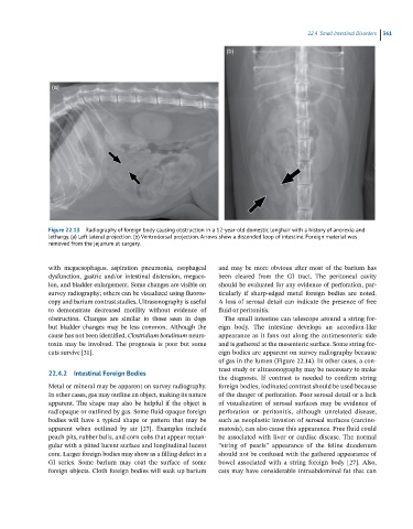
Figure 22.13 Radiography of foreign body causing obstruction in a 12-year-old domestic longhair with a history of anorexia and
lethargy. (a) Left lateral projection. (b) Ventrodorsal projection. Arrows show a distended loop of intestine. Foreign material was
removed from the jejunum at surgery.
with megaesophagus, aspiration pneumonia, esophageal and may be more obvious after most of the barium has
dysfunction, gastric and/or intestinal distension, megaco- been cleared from the GI tract. The peritoneal cavity
lon, and bladder enlargement. Some changes are visible on should be evaluated for any evidence of perforation, par-
survey radiography; others can be visualized using fluoros- ticularly if sharp‐edged metal foreign bodies are noted.
copy and barium contrast studies. Ultrasonography is useful A loss of serosal detail can indicate the presence of free
to demonstrate decreased motility without evidence of fluid or peritonitis.
obstruction. Changes are similar to those seen in dogs The small intestine can telescope around a string for-
but bladder changes may be less common. Although the eign body. The intestine develops an accordion‐like
cause has not been identified, Clostridium botulinum neuro- appearance as it fans out along the antimesenteric side
toxin may be involved. The prognosis is poor but some and is gathered at the mesenteric surface. Some string for-
cats survive [31]. eign bodies are apparent on survey radiography because
of gas in the lumen (Figure 22.14). In other cases, a con-
trast study or ultrasonography may be necessary to make
22.4.2 Intestinal Foreign Bodies
the diagnosis. If contrast is needed to confirm string
Metal or mineral may be apparent on survey radiography. foreign bodies, iodinated contrast should be used because
In other cases, gas may outline an object, making its nature of the danger of perforation. Poor serosal detail or a lack
apparent. The shape may also be helpful if the object is of visualization of serosal surfaces may be evidence of
radiopaque or outlined by gas. Some fluid‐opaque foreign perforation or peritonitis, although unrelated disease,
bodies will have a typical shape or pattern that may be such as neoplastic invasion of serosal surfaces (carcino-
apparent when outlined by air [27]. Examples include matosis), can also cause this appearance. Free fluid could
peach pits, rubber balls, and corn cobs that appear rectan- be associated with liver or cardiac disease. The normal
gular with a pitted lucent surface and longitudinal lucent “string of pearls” appearance of the feline duodenum
core. Larger foreign bodies may show as a filling defect in a should not be confused with the gathered appearance of
GI series. Some barium may coat the surface of some bowel associated with a string foreign body [27]. Also,
foreign objects. Cloth foreign bodies will soak up barium cats may have considerable intraabdominal fat that can