
Page 357 - Feline diagnostic imaging
P. 357
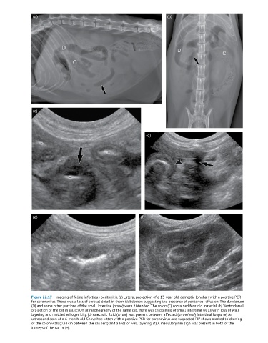
(a)                                                          (b)


                (c)


                                                                    (d)


                (e)                                              (f)


               Figure 22.17  Imaging of feline infectious peritonitis. (a) Lateral projection of a 13-year-old domestic longhair with a positive PCR
               for coronavirus. There was a loss of serosal detail in the midabdomen suggesting the presence of peritoneal effusion. The duodenum
               (D) and some other portions of the small intestine (arrow) were distended. The colon (C) contained fecaloid material. (b) Ventrodorsal
               projection of the cat in (a). (c) On ultrasonography of the same cat, there was thickening of small intestinal walls with loss of wall
               layering and mottled echogenicity. (d) Anechoic fluid (arrow) was present between affected (arrowhead) intestinal loops. (e) An
               ultrasound scan of a 6-month-old Snowshoe kitten with a positive PCR for coronavirus and suspected FIP shows marked thickening
               of the colon wall (0.33 cm between the calipers) and a loss of wall layering. (f) A medullary rim sign was present in both of the
               kidneys of the cat in (e).