
Page 361 - Feline diagnostic imaging
P. 361
(a)
(b)
(c) (d)
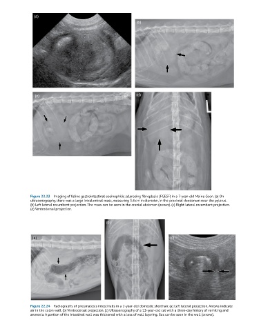
Figure 22.23 Imaging of feline gastrointestinal eosinophilic sclerosing fibroplasia (FGESF) in a 7-year-old Maine Coon. (a) On
ultrasonography, there was a large intraluminal mass, measuring 3.4 cm in diameter, in the proximal duodenum near the pylorus.
(b) Left lateral recumbent projection. The mass can be seen in the cranial abdomen (arrows). (c) Right lateral recumbent projection.
(d) Ventrodorsal projection.
(b)
(a) (c)
Figure 22.24 Radiographs of pneumatosis intestinalis in a 2-year-old domestic shorthair. (a) Left lateral projection. Arrows indicate
air in the colon wall. (b) Ventrodorsal projection. (c) Ultrasonography of a 12-year-old cat with a three-day history of vomiting and
anorexia. A portion of the intestinal wall was thickened with a loss of wall layering. Gas can be seen in the wall (arrows).