
Page 363 - Feline diagnostic imaging
P. 363
22.4 Small ntestinal Disorders 371
(a) (b)
(c) (d)
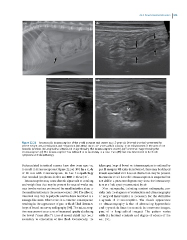
Figure 22.26 Ileocecocolic intussusception of the small intestine and cecum in a 15-year-old Oriental shorthair presented for
severe weight loss, constipation, and megacolon. (a) Lateral projection shows a fluid opacity in the midabdomen in the area of the
ileocolic junction. (b) Longitudinal ultrasound image showing the intussusceptum (arrows). (c) Transverse image showing the
intussusceptum. (d) The intussusception was believed to be secondary to a cecal mass (M) that was determined to be B cell
lymphoma at histopathology.
Pedunculated intestinal masses have also been reported telescoped loop of bowel or intussusceptum is outlined by
to result in intussusception (Figure 22.26) [49]. In a study gas. If an upper GI series is performed, there may be delayed
of 20 cats with intussusception, 16 had histopathology transit associated with ileus or obstruction may be present.
that revealed lymphoma in five and IBD in three [50]. In cases in which ileocolic intussusception is suspected but
Intussusception may cause chronic signs such as vomiting not visible, a pneumocologram may show the intussuscep-
and weight loss that may be present for several weeks and tum as a fluid opacity surrounded by air.
may involve various portions of the small intestine alone or Often radiography, including contrast radiography, pro-
the small intestine into the colon or cecum [50]. The affected vides only the diagnosis of obstruction and ultrasonography
intestinal loop may be palpable and has been described as a or surgical intervention is necessary for the definitive
sausage‐like mass. Obstruction is a common consequence, diagnosis of intussusception. The classic appearance
resulting in the appearance of gas‐ or fluid‐filled distended on ultrasonography is that of alternating hyperechoic
loops of bowel on survey radiography [50]. The intussuscep- and hypoechoic lines (concentric in transverse images,
tion may present as an area of increased opacity displacing parallel in longitudinal images). The pattern varies
the bowel (“mass effect”). Loss of serosal detail may occur with the luminal contents and degree of edema of the
secondary to emaciation or free fluid. Occasionally, the wall [50].