
Page 359 - Feline diagnostic imaging
P. 359
22.4 Small ntestinal Disorders 367
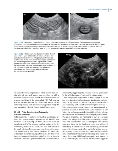
(a) (b)
Figure 22.20 Ultrasound image of mast cell tumor in the small intestine of a 17-year-old cat. The spleen was enlarged on
radiographs. On ultrasound, in addition to splenomegaly, there was thickening of small intestinal loops and enlargement of lymph
nodes. Aspirates of the spleen and lymph nodes revealed mast cell tumor. (a) A longitudinal scan shows thickening of the small
intestine, particularly the muscularis layer. (b) In this transverse image, the muscularis is almost anechoic.
Figure 22.21 Ultrasonography of gastrointestinal stromal
tumor in a 14-year-old domestic shorthair. A mass in the
cranial abdomen had mixed echogenicity and measured
4.06 × 2.27 cm in one plane. The mass was poorly exfoliative
on ultrasound-guided fine needle aspiration but a few
spindle cells were seen. On exploratory laparotomy, there
was a mass near the distal portion of the right extremity of
the pancreas. Portions of the descending duodenum,
ascending colon, ileum, and cecum were adhered.
Histopathology revealed GIST.
echogenicity. Some neoplasms or other lesions may con- lesions [45], suggesting that bacteria or other agents may
tain mineral. Mast cell tumors were usually focal with a be the initiating cause of eosinophilic inflammation.
thickened hypoechoic wall, which was noncircumferential The sonographic appearance of FGESF (Figure 22.23)
in about two‐thirds of the cats studied [43]. Wall layering has been described in the stomach, duodenum, and jeju-
was lost in two‐thirds of the masses and altered in the num [19,39]. In one cat, a focal 2 cm jejunal lesion exhib-
remaining masses, with the muscularis propria being the ited thickening and altered wall layering that resulted in
layer most often affected. One cat had diffuse change. luminal narrowing. Partial obstruction was evidenced by
proximal dilation of the intestine. After resection of the
22.4.4.2 Feline Gastrointestinal Eosinophilic lesion, the cat later presented with additional lesions in the
Sclerosing Fibroplasia small intestine and stomach and enlarged jejunal nodes.
Following review of archived postmortem and surgical tis- The colon of another cat was found to have a 6 cm mass
sues, the histopathologic appearance of FGESF was with mixed echogenicity. The mass contained hyperechoic
described in 25 cats [38]. Of these, 12 had an ulcerated areas and one anechoic area. An echogenic intraluminal
mass in the wall of the pylorus, nine had lesions at the ile- mass was found in the duodenum in an area with wall
ocecocolic junction or the colon, and four had lesions in thickening. A fourth cat had wall thickening in variable
the small intestine. Lymph nodes were abnormal in seven areas of the jejunum and colon, particularly the muscula-
cats. Histologically, the lesions consisted of fibroblasts, ris. A poorly echogenic mass that contained hyperechoic
eosinophils, and dense collagen trabeculae. Bacteria were areas was found in the jejunum extending from an area of
found at the center of the lesion in 14 of the 25 cats. Bacteria mural thickening. Mild lymphadenopathy was present in
were also found in a previous study of cats with similar all of these cats [39]. As noted in the discussion of pyloric