
Page 358 - Feline diagnostic imaging
P. 358
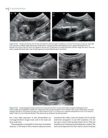
366  22  Gastrointestinal Disease

             (a)                                             (b)


            Figure 22.18  Ultrasound images of a 19-year-old domestic shorthair with intermittent vomiting. The cat had an ileocolic mass that
            was confirmed as diffuse large centroblastic lymphoma at necropsy. (a) The small intestinal wall is greatly thickened at 0.5 cm
            between the calipers. No wall layers are apparent and the wall is decreased in echogenicity. (b) In another image, the small intestinal
            wall is greatly thickened with a loss of wall layering. An arrow indicates gas in the lumen.


                       (a)                                          (b)


                                       (c)


            Figure 22.19  Ultrasonography of adenocarcinoma in the jejunum of an 11-year-old domestic longhair. Radiographs were
            unremarkable. (a) A longitudinal ultrasound image shows the wall thickened at 0.594 cm between the calipers. Wall layers cannot be
            distinguished. (b) In this transverse image, the wall measured 0.532 cm. (c) There was moderate enlargement of one of the adjacent
            lymph nodes. An arrow indicates the tip of the needle during ultrasound-guided aspiration.
            has  a  fixed  rigid  appearance  so  that  abnormalities  are   mentioned above [42], a mass was found in all 16 cats that
            unchanged between images made early in the study and   underwent sonography. In cats with lymphoma [17], the
            those made later.                                 key sign is a loss of wall layering (Figure 22.22). Often the
              Ultrasonography is invaluable for detection of intestinal   intestinal wall is thickened and echogenicity is altered. The
            neoplasia. In the study of feline intestinal adenocarcinoma   wall may be hypoechoic but there may be areas of increased