
Page 356 - Feline diagnostic imaging
P. 356
364 22 Gastrointestinal Disease
(a) (b) (c)
(d) (e) (f)
(g) (h) (i)
(j)
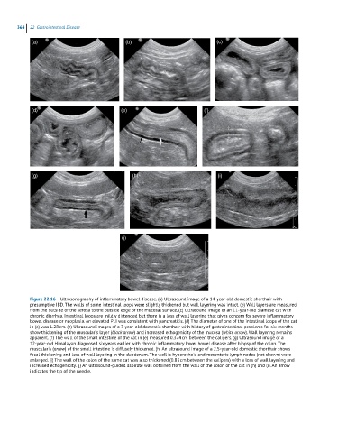
Figure 22.16 Ultrasonography of inflammatory bowel disease. (a) Ultrasound image of a 14-year-old domestic shorthair with
presumptive IBD. The walls of some intestinal loops were slightly thickened but wall layering was intact. (b) Wall layers are measured
from the outside of the serosa to the outside edge of the mucosal surface. (c) Ultrasound image of an 11-year-old Siamese cat with
chronic diarrhea. Intestinal loops are mildly distended but there is a loss of wall layering that gives concern for severe inflammatory
bowel disease or neoplasia. An elevated PLI was consistent with pancreatitis. (d) The diameter of one of the intestinal loops of the cat
in (c) was 1.28 cm. (e) Ultrasound images of a 7-year-old domestic shorthair with history of gastrointestinal problems for six months
show thickening of the muscularis layer (black arrow) and increased echogenicity of the mucosa (white arrow). Wall layering remains
apparent. (f) The wall of the small intestine of the cat in (e) measured 0.374 cm between the calipers. (g) Ultrasound image of a
12-year-old Himalayan diagnosed six years earlier with chronic inflammatory lower bowel disease after biopsy of the colon. The
muscularis (arrow) of the small intestine is diffusely thickened. (h) An ultrasound image of a 2.5-year-old domestic shorthair shows
focal thickening and loss of wall layering in the duodenum. The wall is hyperechoic and mesenteric lymph nodes (not shown) were
enlarged. (i) The wall of the colon of the same cat was also thickened (0.83 cm between the calipers) with a loss of wall layering and
increased echogenicity. (j) An ultrasound-guided aspirate was obtained from the wall of the colon of the cat in (h) and (i). An arrow
indicates the tip of the needle.