
Page 365 - Feline diagnostic imaging
P. 365
22.5 Large ntestinal Disorders 373
22.5 Large Intestinal Disorders intussusception but the intussuscepted loop (intussuscipi-
ens) tends to be smaller in cecal inversion than ileocolic
22.5.1 Cecal Inversion intussusception. Pneumocolo graphy or ultrasonography
could be done for further evaluation.
Cecal inversion is much less common but has been reported
in a cat with ileocolic intussusception [52]. After ultra- 22.5.2 Ileocecocolic Abnormalities
sound revealed a blind sac within the ascending colon, an
ileocolic intussusception and cecal inversion were found at Changes in the ileocecocolic area (Figure 22.28) were
surgery. Gas is not normally seen in the feline cecum, associated with acute diarrhea and vomiting in a study of
unlike in the canine cecum. In dogs, the radiographic 29 cats [53]. Seven cats had a fluid‐filled cecum with one
appearance of cecal inversion is similar to that of ileocolic cat also having material within the cecum that exhibited
(a) (c)
(b)
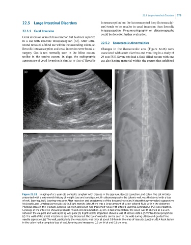
Figure 22.28 Imaging of a 5-year-old domestic longhair with disease in the jejunum, ileocolic junction, and colon. The cat initially
presented with a one-month history of weight loss and constipation. On ultrasonography, the colonic wall was thickened with a loss
of wall layering. Wall layering was poor. After resection and anastomosis of the descending colon, histopathology revealed suppurative,
histiocytic, and lymphoplasmocytic colitis. Eight months later, there was a large amount of straw-colored fluid within the abdomen.
Multiple areas in the jejunum, ileocolic junction, and colon had thickened walls with altered layering. Coronavirus PCR was negative.
Cytology of the intestine showed probable mixed cell inflammation. (a) On initial presentation, the colon was thickened at 0.63 cm
between the calipers and wall layering was poor. (b) Right lateral projection shows a loss of serosal detail. (c) Ventrodorsal projection.
(d) The wall of the small intestine is severely thickened. The tip of a needle can be seen in the wall during ultrasound-guided fine
needle aspiration. (e) The wall, particularly the muscularis, was thick at about 0.84 cm in the area of ileocolic junction. (f) A focal lesion
in the colon had a complete loss of wall layering and measured 0.6 cm thick and 0.8 cm long.