
Page 283 - Veterinary Immunology, 10th Edition
P. 283
Once they have captured and processed antigens, immature DCs
VetBooks.ir carry these antigens to sites where they can be recognized by T
cells. The activated DCs are attracted to lymphoid organs by
chemokines. Infection or tissue damage also promotes the
migration of antigen-bearing DCs to lymph nodes or the spleen.
Once they enter a lymphoid organ, the cells mature rapidly.
Mature DCs secrete chemokines that attract T cells, which
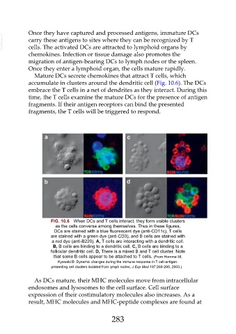
accumulate in clusters around the dendritic cell (Fig. 10.6). The DCs
embrace the T cells in a net of dendrites as they interact. During this
time, the T cells examine the mature DCs for the presence of antigen
fragments. If their antigen receptors can bind the presented
fragments, the T cells will be triggered to respond.
FIG. 10.6 When DCs and T cells interact, they form visible clusters
as the cells converse among themselves. Thus in these figures,
DCs are stained with a blue fluorescent dye (anti-CD11c), T cells
are stained with a green dye (anti-CD3), and B cells are stained with
a red dye (anti-B220). A, T cells are interacting with a dendritic cell.
B, B cells are binding to a dendritic cell. C, B cells are binding to a
follicular dendritic cell. D, There is a mixed B and T cell cluster. Note
that some B cells appear to be attached to T cells. (From Hommel M,
Kyewski B: Dynamic changes during the immune response in T cell-antigen-
presenting cell clusters isolated from lymph nodes, J Exp Med 197:269-280, 2003.)
As DCs mature, their MHC molecules move from intracellular
endosomes and lysosomes to the cell surface. Cell surface
expression of their costimulatory molecules also increases. As a
result, MHC molecules and MHC-peptide complexes are found at
283