
Page 331 - Veterinary Immunology, 10th Edition
P. 331
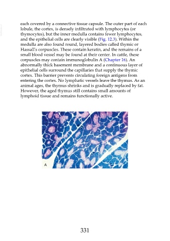
each covered by a connective tissue capsule. The outer part of each
  VetBooks.ir  lobule, the cortex, is densely infiltrated with lymphocytes (or

               thymocytes), but the inner medulla contains fewer lymphocytes,
               and the epithelial cells are clearly visible (Fig. 12.3). Within the

               medulla are also found round, layered bodies called thymic or
               Hassall's corpuscles. These contain keratin, and the remains of a
               small blood vessel may be found at their center. In cattle, these
               corpuscles may contain immunoglobulin A (Chapter 16). An

               abnormally thick basement membrane and a continuous layer of
               epithelial cells surround the capillaries that supply the thymic
               cortex. This barrier prevents circulating foreign antigens from
               entering the cortex. No lymphatic vessels leave the thymus. As an

               animal ages, the thymus shrinks and is gradually replaced by fat.
               However, the aged thymus still contains small amounts of
               lymphoid tissue and remains functionally active.


                                                         331