
Page 437 - Veterinary Immunology, 10th Edition
P. 437
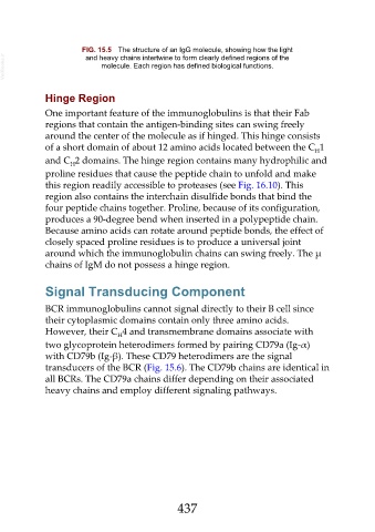
FIG. 15.5  The structure of an IgG molecule, showing how the light
  VetBooks.ir                    molecule. Each region has defined biological functions.
                            and heavy chains intertwine to form clearly defined regions of the


               Hinge Region

               One important feature of the immunoglobulins is that their Fab
               regions that contain the antigen-binding sites can swing freely
               around the center of the molecule as if hinged. This hinge consists
               of a short domain of about 12 amino acids located between the C 1
                                                                                                     H
               and C 2 domains. The hinge region contains many hydrophilic and
                       H
               proline residues that cause the peptide chain to unfold and make
               this region readily accessible to proteases (see Fig. 16.10). This
               region also contains the interchain disulfide bonds that bind the
               four peptide chains together. Proline, because of its configuration,

               produces a 90-degree bend when inserted in a polypeptide chain.
               Because amino acids can rotate around peptide bonds, the effect of
               closely spaced proline residues is to produce a universal joint
               around which the immunoglobulin chains can swing freely. The µ

               chains of IgM do not possess a hinge region.


               Signal Transducing Component

               BCR immunoglobulins cannot signal directly to their B cell since
               their cytoplasmic domains contain only three amino acids.

               However, their C 4 and transmembrane domains associate with
                                      H
               two glycoprotein heterodimers formed by pairing CD79a (Ig-α)

               with CD79b (Ig-β). These CD79 heterodimers are the signal
               transducers of the BCR (Fig. 15.6). The CD79b chains are identical in
               all BCRs. The CD79a chains differ depending on their associated
               heavy chains and employ different signaling pathways.


                                                         437