
Page 438 - Veterinary Immunology, 10th Edition
P. 438
VetBooks.ir
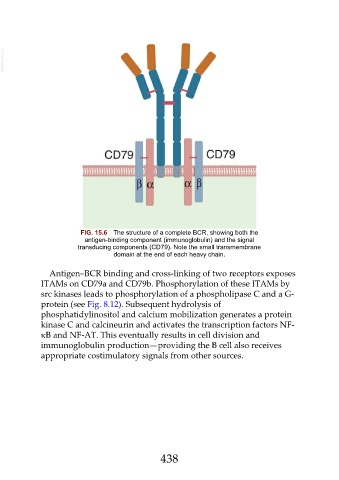
FIG. 15.6 The structure of a complete BCR, showing both the
antigen-binding component (immunoglobulin) and the signal
transducing components (CD79). Note the small transmembrane
domain at the end of each heavy chain.
Antigen–BCR binding and cross-linking of two receptors exposes
ITAMs on CD79a and CD79b. Phosphorylation of these ITAMs by
src kinases leads to phosphorylation of a phospholipase C and a G-
protein (see Fig. 8.12). Subsequent hydrolysis of
phosphatidylinositol and calcium mobilization generates a protein
kinase C and calcineurin and activates the transcription factors NF-
κB and NF-AT. This eventually results in cell division and
immunoglobulin production—providing the B cell also receives
appropriate costimulatory signals from other sources.
438