
Page 490 - Veterinary Immunology, 10th Edition
P. 490
VetBooks.ir
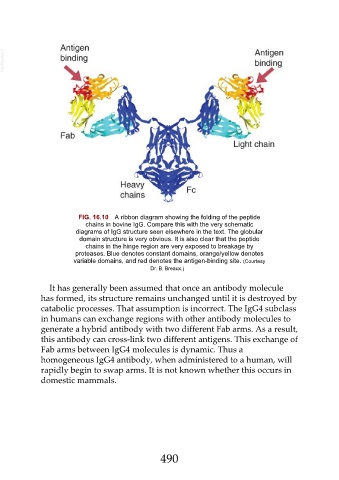
FIG. 16.10 A ribbon diagram showing the folding of the peptide
chains in bovine IgG. Compare this with the very schematic
diagrams of IgG structure seen elsewhere in the text. The globular
domain structure is very obvious. It is also clear that the peptide
chains in the hinge region are very exposed to breakage by
proteases. Blue denotes constant domains, orange/yellow denotes
variable domains, and red denotes the antigen-binding site. (Courtesy
Dr. B. Breaux.)
It has generally been assumed that once an antibody molecule
has formed, its structure remains unchanged until it is destroyed by
catabolic processes. That assumption is incorrect. The IgG4 subclass
in humans can exchange regions with other antibody molecules to
generate a hybrid antibody with two different Fab arms. As a result,
this antibody can cross-link two different antigens. This exchange of
Fab arms between IgG4 molecules is dynamic. Thus a
homogeneous IgG4 antibody, when administered to a human, will
rapidly begin to swap arms. It is not known whether this occurs in
domestic mammals.
490