Page 167 - Manual of Equine Field Surgery
P. 167
Nasolacrimal Flush 163
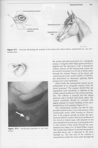
--- Dorsal lacrimal punctum
Nasolacrimal sac --+
'
0\
'------Ventral lacrimal
punctum
Figure 27-1 Schematic illustrating the anatomy of the dorsal and ventral puncta, nasolacrimal sac, and naso-
lacrimal duct.
the ostium and advanced gently in a retrograde
manner.1 Using the index finger, gentle pressure is
applied over the opening to "seal" it around the
catheter. Patency of the nasolacrimal system can
be tested by injecting 15 to 20 mL of sterile saline
through the catheter. Patency of the dorsal and
ventral puncta at the medial canthus is individu-
ally determined by alternately applying digital
pressure over each canaliculus.
Alternatively, the nasolacrimal system can be
catheterized in an antegrade manner from the
dorsal punctum." The surgeon should first use
appropriate local anesthesia or akinesia of the
eyelids, topical anesthesia of the cornea, and seda-
tion. The catheter is inserted into the dorsal
punctum and is flushed with sterile saline. The
ventral canaliculus should be occluded with
digital pressure to ensure flushing of the naso-
lacrimal duct to its opening (Figure 27-3).
The nasolacrimal catheter can also be tem-
porarily sutured in place to provide patency of the
nasolacrimal system while treating an obstruc-
tion. If the surgeon intends to suture the naso-
lacrimal catheter in place, then local anesthesia of
the dorsal or lateral nasal wall adjacent to the
nares should be injected prior to placing the tube.
A stab incision is then made through the anes-
thetized area using a No. 10 Bard-Parker scalpel.
Figure 27-2 Nasolacrimal punctum in the vesti- The nasolacrimal catheter is inserted retrograde as
bulum.
described above and is advanced to the naso-
lacrimal sac. The free end of the catheter is
inserted from inside the nasal cavity through the

