
Page 150 - Veterinary Histology of Domestic Mammals and Birds, 5th Edition
P. 150
132 Veterinary Histology of Domestic Mammals and Birds
VetBooks.ir
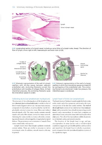
6.22 Longitudinal section of a lymph vessel, including a valve (hilus of a lymph node, sheep). The direction of
flow of lymph is from right to left. Haematoxylin and eosin stain (x120).
Bundle of
collagen fibrils
Access to
intercellular space
Endothelium of
lymph capillary
Anchoring
filament
6.23 Schematic representation of the wall of a lymph 6.24 Schematic representation of the wall of a lymph
capillary with slit-like spaces between adjacent capillary in which the intercellular spaces are closed off
endothelial cells. Anchoring filaments connect the by overlapping of the endothelial cells. The anchor-
endothelium with bundles of collagen fibres. At high ing filaments are shortened. Under low pressure, the
interstitial pressures, the anchoring filaments cause interstitial space is condensed.
the intercellular spaces to widen.
Collecting ducts (vas lymphaticum myotypicum) Lymph heart of birds (cor lymphaticum)
The structure of the collecting ducts of the lymphatic sys- The lymph hearts of birds are located outside the body cavity,
tem (thoracic duct and tracheal trunk) is similar to that of at the caudal end of the synsacrum and dorsal to the trans-
blood vessels (Figures 6.20 to 6.22). An inner tunica intima, verse process of the first free caudal vertebra. The wall of
composed of endothelium, is surrounded by mainly lon- the lymph heart consists of an endothelium-lined endocar-
gitudinally oriented connective tissue fibres and isolated dium (with a connective tissue sheath incorporating smooth
smooth muscle cells. The tunica media contains smooth muscle) and a myocardium. Its outer layer comprises an
muscle cells and a fibro-elastic connective tissue network. adventitia of loose connective tissue with multilocular white
Adjoining the tunica media is a tunica adventitia contain- adipocytes. The cells of the myocardium exhibit characteris-
ing smooth muscle cells arranged in a longitudinal or spiral tics of skeletal, cardiac and smooth muscles.
fashion. The prominence and orientation of muscle fibres The function of the lymph heart is age- and spe-
varies considerably with species. cies-dependent. In avian species with a well-developed
Lymph vessels other than lymph capillaries routinely copulatory organ and a protrusible phallus, such as ratites
contain valves. These are similar in structure to valves and waterfowl, the lymph heart is wholly or predomi-
occurring in veins. In addition, smooth muscle cells may nantly integrated into the specialised lymphatic system of
be present. the copulatory organ.
Vet Histology.indb 132 16/07/2019 14:58