
Page 202 - Veterinary Histology of Domestic Mammals and Birds, 5th Edition
P. 202
184 Veterinary Histology of Domestic Mammals and Birds
Tongue (lingua) Ruminants: A raised area with a thickened mucosa, the
VetBooks.ir The tongue of domestic mammals is a highly mobile torus linguae, is present on the dorsal aspect of the
muscle. The body (corpus linguae) and root (radix
caudal portion of the tongue.
linguae) project into the oral cavity and the tip (apex lin-
guae) moves freely within the cavum oris. The tongue has Birds: The tongue of birds conforms to the shape of the
several functions, including prehension of solid and liquid lower beak. In the chicken, a transverse row of caudally
nutrients and detection of tactile and sensory stimuli. It is directed papillae lies between the body and the root of
also used as an aid in grooming of the hair coat. the tongue. The body of the tongue is supported by a
The non-glandular lingual mucosa is lined with strati- bone, the paraglossum (= entoglossum), the intrinsic
fied squamous epithelium of varying thickness. In keeping musculature being only rudimentary. The shape and
with the mechanical forces imposed by solid foodstuffs, the development of the avian tongue varies according to
epithelium of the dorsum of the tongue is thick, heavily diet. The tongue of kolibris (hummingbirds) and insec-
keratinised and strongly papillated, particularly in herbi- tivorous species is very long and highly protrusible. In
vores and cats. The margins and ventrum of the tongue are most avian species, including the chicken, the tongue is
lined by a thin stratified squamous epithelium. pointed apically and slightly broadened at its base. The
The considerable mobility of the tongue arises from tongue is lined by non-glandular mucosa, in which the
the three-dimensional arrangement of the striated mus- epithelium exhibits local areas of keratinisation. Ventrally,
cle fibre bundles within the m. lingualis proprius (Figure the tongue is supported by a keratinised plate (cuticula
10.6). The muscle fibres are oriented in three directions cornea lingualis). In ducks and geese, the edges of the
(longitudinal, vertical and transverse), crossing each tongue are lined with spiny keratinised bristles that are
other at right angles to form a regular lattice. The mus- directed towards the pharynx. These assist with selective
cle bundles are separated by connective tissue septa. filtration during feeding on vegetable matter.
Depending on species, these may contain adipose tissue.
Numerous blood vessels and nerves are also present. The Lingual papillae
tongue of the pig is particularly rich in unilocular adipose The surface of the tongue (mostly the dorsum) is covered
tissue. with mucosal elevations that facilitate the uptake and sen-
The lyssa is an unpaired, rod-like support structure sory assessment of food. Termed lingual papillae (papillae
located ventrally at the apex of the tongue. It encloses linguales), these structures vary in their size, number and
adipose tissue, skeletal muscle and connective tissue. The distribution over the surface of the tongue (Figures 10.6 to
lyssa is well developed in carnivores, predominantly filled 10.9). A distinction is made between mechanical papillae
with fat in pigs and rudimentary in horses. (papillae mechanicae) and gustatory papillae (papillae
gustatoriae), which are further subdivided as follows:
Species variation · mechanical papillae (papillae mechanicae):
Horse: A fibro-elastic cord incorporating hyaline carti- − filiform papillae (papillae filiformes),
lage, muscle and adipose tissue (dorsal lingual cartilage) − conical papillae (papillae conicae),
is embedded in the dorsum of the tongue. − marginal papillae (papillae marginales),
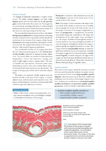
Stratified squamous epithelium
with mechanical papillae
Lamina propria mucosae
Intrinsic muscle of the tongue with
horizontally, vertically and longitudinally
oriented fibres
Lyssa
10.6 Cross-section of the tongue (dog).
The dorsum of the tongue is covered
with numerous mechanical papillae and
is distinctly papillated. The orientation
of the muscle fibres forms a three-
dimensional lattice. The tongue of the dog
is supported by the centrally located lyssa.
Goldner’s Masson trichrome stain (x20).
Vet Histology.indb 184 16/07/2019 15:00