
Page 200 - Veterinary Histology of Domestic Mammals and Birds, 5th Edition
P. 200
182 Veterinary Histology of Domestic Mammals and Birds
Cheek (bucca) Palate (palatum)
VetBooks.ir The cheeks (Figure 10.4) are similar in structure to the lips, The palate comprises the hard palate (palatum durum),
which covers the bones of the roof of the oral cavity, and
comprising the following layers:
the soft palate (palatum molle, velum palatinum), which
· outer skin layer, extends towards the pharynx.
· middle muscle layer and
· inner mucosal layer.
Hard palate (palatum durum)
Unlike the lips, the cheeks are covered externally by hair. The lining of the hard palate comprises:
The middle layer consists of the buccal musculature. The
inner wall is composed of non-glandular mucosa lined by · keratinised stratified squamous epithelium that
stratified squamous epithelium. In ruminants, buccal papil- interdigitates with collagen-rich subepithelial con-
lae (papillae conicae buccales) are present on the surface nective tissue (papillation) and
of the mucosa. · deeper connective tissue containing small vessels
The tela submucosa contains extensive aggregates of and a large venous plexus, as well as numerous
buccal glands (glandulae buccales) that distribute their fibrocytes, lymphocytes and macrophages (fuses
secretory product to the vestibulum oris through short with subepithelial connective tissue to form a
ducts. The buccal glands are minor salivary glands. They propria-submucosa).
extend deep into the buccal musculature, which aids in
expressing the secretory product during chewing. The The mucosa of the hard palate is lined by stratified squa-
extent of the buccal glands and the composition of their mous epithelium. It is a mostly non-glandular, tough
secretions varies with species. Morphologically, they layer of tissue that is tightly bound to the periosteum of
are classified as compound tubulo-acinar glands. Based the underlying bone. The mucosa exhibits transverse
on their secretory product, buccal glands are divided ridges (rugae palatinae) that vary in number and promi-
into: nence, depending upon species. The surface of the ridges
is reinforced by thick sheets of non-glandular, heavily
· serous glands: ventral buccal glands of cattle, keratinised epithelium. Isolated mixed glands (glandu-
· mucous glands: middle and dorsal buccal glands lae parapapillares) occur between the caudal ridges in
of cattle, ventral buccal glands of sheep, goats and ruminants and in the dog, and more rostrally in the pig.
carnivores and The taut lamina propria blends without obvious demarca-
· mixed glands: buccal glands of horses and pigs. tion with the tela submucosa, through which it is tightly
fused to the periosteum. In carnivores and (particularly)
in ungulates, the propria-submucosa contains an extensive
branching venous network. In ruminants, the mucosa is
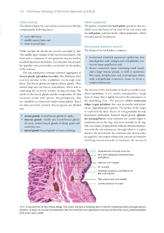
10.4 Cross-section of the cheek (dog). The outer surface is lined by skin in which numerous hairs and glands are
present. A layer of muscle lies between the skin and the non-glandular mucosa of the oral cavity. Haematoxylin
and eosin stain (x26).
Vet Histology.indb 182 16/07/2019 15:00