
Page 196 - Veterinary Histology of Domestic Mammals and Birds, 5th Edition
P. 196
178 Veterinary Histology of Domestic Mammals and Birds
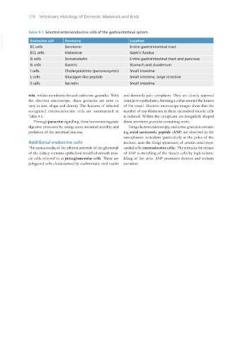
Table 9.1 Selected enteroendocrine cells of the gastrointestinal system.
VetBooks.ir Endocrine cell Hormone Location
EC cells
Serotonin
Entire gastrointestinal tract
ECL cells Histamine Gastric fundus
D cells Somatostatin Entire gastrointestinal tract and pancreas
G cells Gastrin Stomach and duodenum
I cells Cholecystokinin (pancreozymin) Small intestine
L cells Glucagon-like peptide Small intestine, large intestine
S cells Secretin Small intestine
tein, within membrane-bound endocrine granules. With and distinctly pale cytoplasm. They are closely apposed
the electron microscope, these granules are seen to (similar to epithelium), forming a collar around the lumen
vary in size, shape and density. The features of selected of the vessel. Electron microscope images show that the
recognised enteroendocrine cells are summarised in number of myofilaments in these specialised muscle cells
Table 9.1. is reduced. Within the cytoplasm are irregularly shaped
Through paracrine signalling, these hormones regulate dense secretory granules containing renin.
digestive processes by acting upon intestinal motility and Using electron microscopy, endocrine granules contain-
perfusion of the intestinal mucosa. ing atrial natriuretic peptide (ANP) are observed in the
sarcoplasmic reticulum (particularly at the poles of the
Additional endocrine cells nucleus, near the Golgi apparatus) of certain atrial myo-
The tunica media of the afferent arteriole of the glomeruli cardial cells (myoendocrine cells). The stimulus for release
of the kidney contains epithelioid modified smooth mus- of ANP is stretching of the muscle cells by high-volume
cle cells referred to as juxtaglomerular cells. These are filling of the atria. ANP promotes diuresis and sodium
polygonal cells characterised by euchromatic oval nuclei excretion.
Vet Histology.indb 178 16/07/2019 15:00